


Mechanistic insight from the crystal structure of A.aolicus LpxC in the presence of product
Miller, M.D., Gao, N., Ross, P., Olivier, N.B.To be published.
Experimental Data Snapshot
Starting Model: experimental
View more details
Entity ID: 1 | |||||
|---|---|---|---|---|---|
| Molecule | Chains | Sequence Length | Organism | Details | Image |
| UDP-3-O-[3-hydroxymyristoyl] N-acetylglucosamine deacetylase | 282 | Aquifex aeolicus | Mutation(s): 0 Gene Names: lpxC, envA, aq_1772 EC: 3.5.1 (PDB Primary Data), 3.5.1.108 (UniProt) | 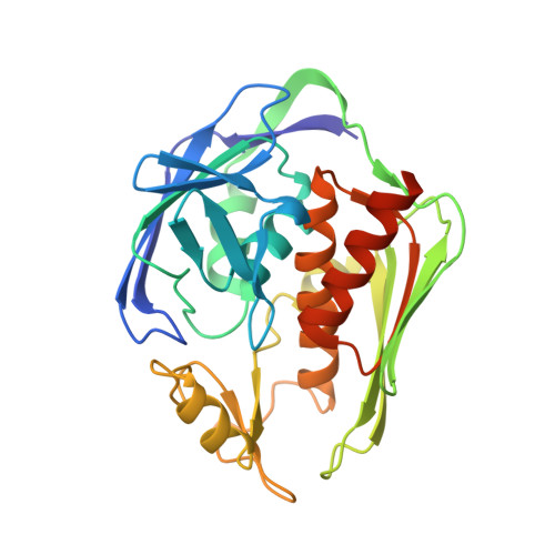 | |
UniProt | |||||
Entity Groups | |||||
| Sequence Clusters | 30% Identity50% Identity70% Identity90% Identity95% Identity100% Identity | ||||
| UniProt Group | O67648 | ||||
Sequence AnnotationsExpand | |||||
Reference Sequence | |||||
| Ligands 3 Unique | |||||
|---|---|---|---|---|---|
| ID | Chains | Name / Formula / InChI Key | 2D Diagram | 3D Interactions | |
| 24G Download:Ideal Coordinates CCD File | H [auth A], M [auth B] | uridine-5'-diphosphate-3-O-(R-3-hydroxymyristoyl)-glucosamine C29 H51 N3 O18 P2 ZFPNNOXCEDQJQS-SSVOXRMNSA-N |  | ||
| ZN Download:Ideal Coordinates CCD File | C [auth A] D [auth A] E [auth A] F [auth A] I [auth B] | ZINC ION Zn PTFCDOFLOPIGGS-UHFFFAOYSA-N |  | ||
| CL Download:Ideal Coordinates CCD File | G [auth A], L [auth B] | CHLORIDE ION Cl VEXZGXHMUGYJMC-UHFFFAOYSA-M |  | ||
| Length ( Å ) | Angle ( ˚ ) |
|---|---|
| a = 101.332 | α = 90 |
| b = 101.332 | β = 90 |
| c = 123.553 | γ = 120 |
| Software Name | Purpose |
|---|---|
| BUSTER | refinement |
| PHASER | phasing |
| PDB_EXTRACT | data extraction |


