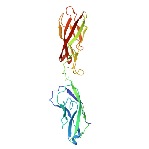
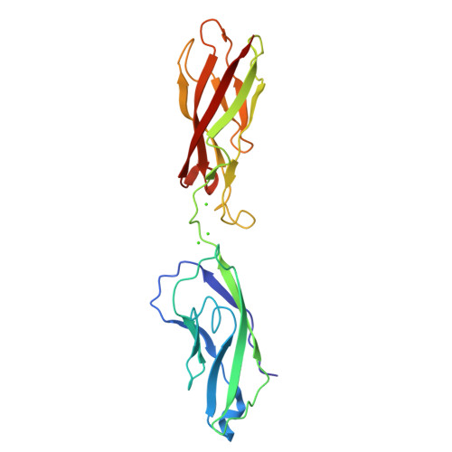
The X-ray structure of human P-cadherin EC1-EC2 in a closed conformation provides insight into the type I cadherin dimerization pathway.
Dalle Vedove, A., Lucarelli, A.P., Nardone, V., Matino, A., Parisini, E.(2015) Acta Crystallogr F Struct Biol Commun 71: 371-380
- PubMed: 25849494
- DOI: https://doi.org/10.1107/S2053230X15003878
- Primary Citation of Related Structures:
4OY9 - PubMed Abstract:
Cadherins are a large family of calcium-dependent proteins that mediate cellular adherens junction formation and tissue morphogenesis. To date, the most studied cadherins are those classified as classical, which are further divided into type I or type II depending on selected sequence features. Unlike other members of the classical cadherin family, a detailed structural characterization of P-cadherin has not yet been fully obtained. Here, the high-resolution crystal structure determination of the closed form of human P-cadherin EC1-EC2 is reported. The structure shows a novel, monomeric packing arrangement that provides a further snapshot in the yet-to-be-achieved complete description of the highly dynamic cadherin dimerization pathway. Moreover, this is the first multidomain cadherin fragment to be crystallized and structurally characterized in its closed conformation that does not carry any extra N-terminal residues before the naturally occurring aspartic acid at position 1. Finally, two clear alternate conformations are observed for the critical Trp2 residue, suggestive of a transient, metastable state. The P-cadherin structure and packing arrangement shown here provide new and valuable information towards the complete structural characterization of the still largely elusive cadherin dimerization pathway.
Organizational Affiliation:
Center for Nano Science and Technology @PoliMi, Istituto Italiano di Tecnologia, Via Pascoli 70/3, 20133 Milan, Italy.


