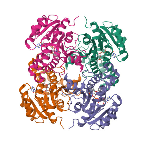
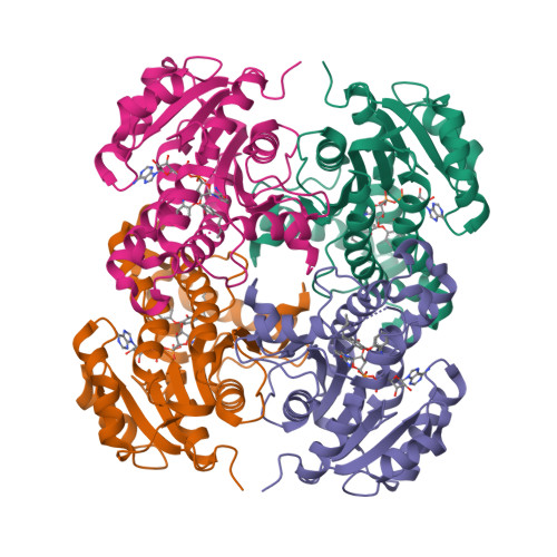
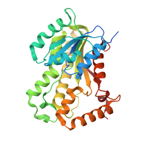
4OXY

Substrate-binding loop movement with inhibitor PT10 in the tetrameric Mycobacterium tuberculosis enoyl-ACP reductase InhA


Experimental Data Snapshot

  • Method: X-RAY DIFFRACTION
  • Resolution: 2.35 Å
  • R-Value Free: 
    0.220 (Depositor), 0.210 (DCC) 
  • R-Value Work: 
    0.166 (Depositor), 0.160 (DCC) 
  • R-Value Observed: 
    0.169 (Depositor) 

Starting Model: experimental
View more details

wwPDB Validation   3D Report Full Report


Ligand Structure Quality Assessment 

Created with Raphaël 2.3.0Worse 01 BetterLigand structure goodness of fit to experimental dataBest fitted NADClick on this verticalbar to view detailsBest fitted 1TNClick on this verticalbar to view details

This is version 1.3 of the entry. See complete history


Literature

A Structural and Energetic Model for the Slow-Onset Inhibition of the Mycobacterium tuberculosis Enoyl-ACP Reductase InhA.

Li, H.J.Lai, C.T.Pan, P.Yu, W.Liu, N.Bommineni, G.R.Garcia-Diaz, M.Simmerling, C.Tonge, P.J.

(2014) ACS Chem Biol 9: 986-993

  • DOI: https://doi.org/10.1021/cb400896g
  • Primary Citation of Related Structures:  
    4OHU, 4OXK, 4OXN, 4OXY, 4OYR

  • PubMed Abstract: 

    Slow-onset enzyme inhibitors are of great interest for drug discovery programs since the slow dissociation of the inhibitor from the drug-target complex results in sustained target occupancy leading to improved pharmacodynamics. However, the structural basis for slow-onset inhibition is often not fully understood, hindering the development of structure-kinetic relationships and the rational optimization of drug-target residence time. Previously we demonstrated that slow-onset inhibition of the Mycobacterium tuberculosis enoyl-ACP reductase InhA correlated with motions of a substrate-binding loop (SBL) near the active site. In the present work, X-ray crystallography and molecular dynamics simulations have been used to map the structural and energetic changes of the SBL that occur upon enzyme inhibition. Helix-6 within the SBL adopts an open conformation when the inhibitor structure or binding kinetics is substrate-like. In contrast, slow-onset inhibition results in large-scale local refolding in which helix-6 adopts a closed conformation not normally populated during substrate turnover. The open and closed conformations of helix-6 are hypothesized to represent the EI and EI* states on the two-step induced-fit reaction coordinate for enzyme inhibition. These two states were used as the end points for nudged elastic band molecular dynamics simulations resulting in two-dimensional potential energy profiles that reveal the barrier between EI and EI*, thus rationalizing the binding kinetics observed with different inhibitors. Our findings indicate that the structural basis for slow-onset kinetics can be understood once the structures of both EI and EI* have been identified, thus providing a starting point for the rational control of enzyme-inhibitor binding kinetics.


  • Organizational Affiliation

    Institute for Chemical Biology and Drug Discovery, ‡Laufer Center for Physical and Quantitative Biology and §Department of Chemistry, ∥Graduate Program in Biochemistry and Structural Biology, and ⊥Department of Pharmacological Sciences, Stony Brook University , Stony Brook, New York 11794, United States.


Macromolecules
Find similar proteins by:  (by identity cutoff)  |  3D Structure
Entity ID: 1
MoleculeChains Sequence LengthOrganismDetailsImage
Enoyl-[acyl-carrier-protein] reductase [NADH]
A, B, C, D
289Mycobacterium tuberculosisMutation(s): 0 
Gene Names: MT1531MTCY277.05Rv1484inhA
EC: 1.3.1.9
UniProt
Find proteins for P9WGR1 (Mycobacterium tuberculosis (strain ATCC 25618 / H37Rv))
Explore P9WGR1 
Go to UniProtKB:  P9WGR1
Entity Groups  
Sequence Clusters30% Identity50% Identity70% Identity90% Identity95% Identity100% Identity
UniProt GroupP9WGR1
Sequence Annotations
Expand
  • Reference Sequence
Small Molecules
Ligands 2 Unique
IDChains Name / Formula / InChI Key2D Diagram3D Interactions
NAD
Query on NAD

Download Ideal Coordinates CCD File 
E [auth A],
G [auth B],
I [auth C],
K [auth D]
NICOTINAMIDE-ADENINE-DINUCLEOTIDE
C21 H27 N7 O14 P2
BAWFJGJZGIEFAR-NNYOXOHSSA-N
1TN
Query on 1TN

Download Ideal Coordinates CCD File 
F [auth A],
H [auth B],
J [auth C],
L [auth D]
5-hexyl-2-(2-nitrophenoxy)phenol
C18 H21 N O4
ASCHWJRXPZBPHV-UHFFFAOYSA-N
Binding Affinity Annotations 
IDSourceBinding Affinity
1TN BindingDB:  4OXY Ki: 129 (nM) from 1 assay(s)
IC50: min: 180, max: 182 (nM) from 2 assay(s)
Experimental Data & Validation

Experimental Data

  • Method: X-RAY DIFFRACTION
  • Resolution: 2.35 Å
  • R-Value Free:  0.220 (Depositor), 0.210 (DCC) 
  • R-Value Work:  0.166 (Depositor), 0.160 (DCC) 
  • R-Value Observed: 0.169 (Depositor) 
Space Group: P 21 21 21
Unit Cell:
Length ( Å )Angle ( ˚ )
a = 88.826α = 90
b = 91.076β = 90
c = 148.911γ = 90
Software Package:
Software NamePurpose
HKL-2000data collection
HKL-2000data scaling
PHENIXrefinement
PDB_EXTRACTdata extraction
CNSrefinement
MOLREPphasing

Structure Validation

View Full Validation Report


Ligand Structure Quality Assessment 

Created with Raphaël 2.3.0Worse 01 BetterLigand structure goodness of fit to experimental dataBest fitted NADClick on this verticalbar to view detailsBest fitted 1TNClick on this verticalbar to view details

Entry History & Funding Information

Deposition Data


Funding OrganizationLocationGrant Number
National Institutes of Health/National Institute of General Medical Sciences (NIH/NIGMS)United StatesGM102864
Department of Energy (DOE, United States)United States--
National Institutes of Health/National Center for Research Resources (NIH/NCRR)United StatesP41RR012408

Revision History  (Full details and data files)

  • Version 1.0: 2014-04-30
    Type: Initial release
  • Version 1.1: 2017-09-06
    Changes: Advisory, Author supporting evidence, Derived calculations, Other, Refinement description, Source and taxonomy, Structure summary
  • Version 1.2: 2019-12-04
    Changes: Author supporting evidence
  • Version 1.3: 2023-09-27
    Changes: Data collection, Database references, Refinement description