4OQO | pdb_00004oqo

Crystal structure of the tryptic generated iron-free C-lobe of bovine Lactoferrin at 2.42 Angstrom resolution


Experimental Data Snapshot

  • Method: X-RAY DIFFRACTION
  • Resolution: 2.42 Å
  • R-Value Free: 
    0.260 (Depositor), 0.266 (DCC) 
  • R-Value Work: 
    0.223 (Depositor), 0.226 (DCC) 
  • R-Value Observed: 
    0.225 (Depositor) 

Starting Model: experimental
View more details

wwPDB Validation 3D Report Full Report

Validation slider image for 4OQO

Ligand Structure Quality Assessment 


This is version 2.2 of the entry. See complete history

Literature

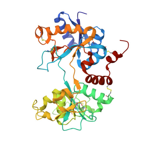
Structure of the iron-free true C-terminal half of bovine lactoferrin produced by tryptic digestion and its functional significance in the gut.

Rastogi, N.Singh, A.Pandey, S.N.Sinha, M.Bhushan, A.Kaur, P.Sharma, S.Singh, T.P.

(2014) FEBS J 281: 2871-2882

  • DOI: https://doi.org/10.1111/febs.12827
  • Primary Citation Related Structures: 
    4OQO

  • PubMed Abstract: 

    Bovine lactoferrin, a 76-kDa glycoprotein (Ala1-Arg689) consists of two similar N- and C-terminal molecular halves with the ability to bind two Fe(3+) ions. The N-terminal half, designated as the N-lobe (Ala1-Arg341) and the C-terminal half designated as the C-lobe (Tyr342-Arg689) have similar iron-binding properties, but the resistant C-lobe prolongs the physiological role of bovine lactoferrin in the digestive tract. Here, we report the crystal structure of true C-lobe, which was produced by limited proteolysis of bovine lactoferrin using trypsin. In the first proteolysis step, two fragments of 21 kDa (Glu86-Lys282) and 45 kDa (Ser283-Arg689) were generated because two lysine residues, Lys85 and Lys282, in the structure of iron-saturated bovine lactoferrin were fully exposed. The 45-kDa fragment was further digested at the newly exposed side chain of Arg341, generating a 38-kDa perfect C-lobe (Tyr342-Arg689). By contrast, the apo-lactoferrin was cut by trypsin only at Arg341, which was exposed in the structure of apo-lactoferrin, whereas the other two sites with Lys85 and Lys282 are inaccessible. The purified iron-saturated C-lobe was crystallized at pH 4.0. The structure was determined by the molecular replacement method using coordinates of the C-terminal half (Arg342-Arg689) of intact camel apo-lactoferrin. The structure determination revealed that the iron atom was absent and the iron-binding cleft was found in a wide-open conformation, whereas in the previously determined structure of iron-saturated C-lobe of bovine lactoferrin, the iron atom was present and the iron-binding site was in the closed confirmation.


  • Organizational Affiliation
    • Department of Biophysics, All India Institute of Medical Sciences, New Delhi, India.

Macromolecule Content 

  • Total Structure Weight: 77.14 kDa 
  • Atom Count: 5,696 
  • Modeled Residue Count: 696 
  • Deposited Residue Count: 696 
  • Unique protein chains: 1

Macromolecules

Find similar proteins by:|  3D Structure
Entity ID: 1
MoleculeChains  Sequence LengthOrganismDetailsImage
Lactotransferrin
A, B
348Bos taurusMutation(s): 0 
EC: 3.4.21
UniProt
Find proteins for P24627 (Bos taurus)
Explore P24627 
Go to UniProtKB:  P24627
Entity Groups
Sequence Clusters30% Identity50% Identity70% Identity90% Identity95% Identity100% Identity
UniProt GroupP24627
Glycosylation
Glycosylation Sites: 2
Sequence Annotations
Expand
Reference Sequence

Oligosaccharides

Help  
Entity ID: 2
MoleculeChains Length2D Diagram GlycosylationD Interactions
2-acetamido-2-deoxy-beta-D-glucopyranose-(1-4)-2-acetamido-2-deoxy-beta-D-glucopyranose
C
2N-Glycosylation
Glycosylation Resources
GlyTouCan: G42666HT
GlyCosmos: G42666HT
GlyGen: G42666HT

Experimental Data & Validation

Experimental Data

  • Method: X-RAY DIFFRACTION
  • Resolution: 2.42 Å
  • R-Value Free:  0.260 (Depositor), 0.266 (DCC) 
  • R-Value Work:  0.223 (Depositor), 0.226 (DCC) 
  • R-Value Observed: 0.225 (Depositor) 
Space Group: P 1 21 1
Unit Cell:
Length ( Å )Angle ( ˚ )
a = 75.81α = 90
b = 49.44β = 94.33
c = 97.86γ = 90
Software Package:
Software NamePurpose
HKL-2000data collection
AMoREphasing
REFMACrefinement
HKL-2000data reduction
SCALEPACKdata scaling

Structure Validation

View Full Validation Report


Ligand Structure Quality Assessment 


Entry History 

Deposition Data

Revision History  (Full details and data files)

  • Version 1.0: 2014-03-12
    Type: Initial release
  • Version 1.1: 2014-07-16
    Changes: Database references
  • Version 2.0: 2020-07-29
    Type: Remediation
    Reason: Carbohydrate remediation
    Changes: Atomic model, Data collection, Derived calculations, Structure summary
  • Version 2.1: 2023-11-08
    Changes: Data collection, Database references, Refinement description, Structure summary
  • Version 2.2: 2024-10-30
    Changes: Structure summary