4OOL | pdb_00004ool

Crystal structure of PBP3 in complex with compound 14 ((2E)-2-({[(2S)-2-{[(2Z)-2-(2-amino-1,3-thiazol-4-yl)-2-{[(1,5-dihydroxy-4-oxo-1,4-dihydropyridin-2-yl)methoxy]imino}acetyl]amino}-3-oxopropyl]oxy}imino)pentanedioic acid)


Experimental Data Snapshot

  • Method: X-RAY DIFFRACTION
  • Resolution: 2.30 Å
  • R-Value Free: 
    0.241 (Depositor), 0.255 (DCC) 
  • R-Value Work: 
    0.186 (Depositor), 0.199 (DCC) 
  • R-Value Observed: 
    0.189 (Depositor) 

Starting Model: experimental
View more details

wwPDB Validation 3D Report Full Report

Validation slider image for 4OOL

Ligand Structure Quality Assessment 


This is version 1.3 of the entry. See complete history

Literature

Siderophore receptor-mediated uptake of lactivicin analogues in gram-negative bacteria.

Starr, J.Brown, M.F.Aschenbrenner, L.Caspers, N.Che, Y.Gerstenberger, B.S.Huband, M.Knafels, J.D.Lemmon, M.M.Li, C.McCurdy, S.P.McElroy, E.Rauckhorst, M.R.Tomaras, A.P.Young, J.A.Zaniewski, R.P.Shanmugasundaram, V.Han, S.

(2014) J Med Chem 57: 3845-3855

  • DOI: https://doi.org/10.1021/jm500219c
  • Primary Citation Related Structures: 
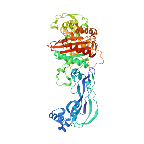
    4OOL, 4OOM, 4OON

  • PubMed Abstract: 

    Multidrug-resistant Gram-negative pathogens are an emerging threat to human health, and addressing this challenge will require development of new antibacterial agents. This can be achieved through an improved molecular understanding of drug-target interactions combined with enhanced delivery of these agents to the site of action. Herein we describe the first application of siderophore receptor-mediated drug uptake of lactivicin analogues as a strategy that enables the development of novel antibacterial agents against clinically relevant Gram-negative bacteria. We report the first crystal structures of several sideromimic conjugated compounds bound to penicillin binding proteins PBP3 and PBP1a from Pseudomonas aeruginosa and characterize the reactivity of lactivicin and β-lactam core structures. Results from drug sensitivity studies with β-lactamase enzymes are presented, as well as a structure-based hypothesis to reduce susceptibility to this enzyme class. Finally, mechanistic studies demonstrating that sideromimic modification alters the drug uptake process are discussed.


  • Organizational Affiliation
    • Medicinal Chemistry, ⧧Computational Chemistry, §Antibacterials Research Unit, and ¶Structural Biology, Pfizer Global Research and Development , Eastern Point Road, Groton, Connecticut 06340, United States.

Macromolecule Content 

  • Total Structure Weight: 58.87 kDa 
  • Atom Count: 3,960 
  • Modeled Residue Count: 499 
  • Deposited Residue Count: 538 
  • Unique protein chains: 1

Macromolecules

Find similar proteins by:|  3D Structure
Entity ID: 1
MoleculeChains  Sequence LengthOrganismDetailsImage
Cell division protein FtsI [Peptidoglycan synthetase]538Pseudomonas aeruginosa PA1RMutation(s): 0 
Gene Names: PA1R_gp2318
EC: 2.4.1.129

Small Molecules

Ligands 1 Unique
IDChains Name / Formula / InChI Key2D Diagram3D Interactions
2U2

Query on 2U2


Download:Ideal Coordinates CCD File
B [auth A](2E)-2-({[(2S)-2-{[(2Z)-2-(2-amino-1,3-thiazol-4-yl)-2-{[(1,5-dihydroxy-4-oxo-1,4-dihydropyridin-2-yl)methoxy]imino}acetyl]amino}-3-oxopropyl]oxy}imino)pentanedioic acid
C19 H20 N6 O11 S
OZMUGJSDFUATCG-ROWSSYOJSA-N

Experimental Data & Validation

Experimental Data

  • Method: X-RAY DIFFRACTION
  • Resolution: 2.30 Å
  • R-Value Free:  0.241 (Depositor), 0.255 (DCC) 
  • R-Value Work:  0.186 (Depositor), 0.199 (DCC) 
  • R-Value Observed: 0.189 (Depositor) 
Space Group: P 21 21 21
Unit Cell:
Length ( Å )Angle ( ˚ )
a = 68.28α = 90
b = 83.54β = 90
c = 89.25γ = 90
Software Package:
Software NamePurpose
autoPROCdata collection
BUSTERrefinement
autoPROCdata scaling
SCALAdata scaling
BUSTERphasing

Structure Validation

View Full Validation Report


Ligand Structure Quality Assessment 


Entry History 

Deposition Data

Revision History  (Full details and data files)

  • Version 1.0: 2014-05-07
    Type: Initial release
  • Version 1.1: 2014-05-21
    Changes: Database references
  • Version 1.2: 2023-09-20
    Changes: Data collection, Database references, Derived calculations, Refinement description
  • Version 1.3: 2024-10-16
    Changes: Structure summary