4ONS | pdb_00004ons

Structural and thermodynamic characterization of cadherin-beta-catenin-alpha-catenin complex formation


Experimental Data Snapshot

  • Method: X-RAY DIFFRACTION
  • Resolution: 2.80 Å
  • R-Value Free: 
    0.264 (Depositor), 0.270 (DCC) 
  • R-Value Work: 
    0.203 (Depositor), 0.211 (DCC) 
  • R-Value Observed: 
    0.207 (Depositor) 

wwPDB Validation 3D Report Full Report

Validation slider image for 4ONS

This is version 1.3 of the entry. See complete history

Literature

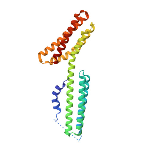
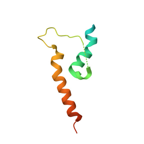
Structural and Thermodynamic Characterization of Cadherin beta-Catenin alpha-Catenin Complex Formation.

Pokutta, S.Choi, H.J.Ahlsen, G.Hansen, S.D.Weis, W.I.

(2014) J Biological Chem 289: 13589-13601

  • DOI: https://doi.org/10.1074/jbc.M114.554709
  • Primary Citation Related Structures: 
    4ONS

  • PubMed Abstract: 

    The classical cadherin·β-catenin·α-catenin complex mediates homophilic cell-cell adhesion and mechanically couples the actin cytoskeletons of adjacent cells. Although α-catenin binds to β-catenin and to F-actin, β-catenin significantly weakens the affinity of α-catenin for F-actin. Moreover, α-catenin self-associates into homodimers that block β-catenin binding. We investigated quantitatively and structurally αE- and αN-catenin dimer formation, their interaction with β-catenin and the cadherin·β-catenin complex, and the effect of the α-catenin actin-binding domain on β-catenin association. The two α-catenin variants differ in their self-association properties: at physiological temperatures, αE-catenin homodimerizes 10× more weakly than does αN-catenin but is kinetically trapped in its oligomeric state. Both αE- and αN-catenin bind to β-catenin with a Kd of 20 nM, and this affinity is increased by an order of magnitude when cadherin is bound to β-catenin. We describe the crystal structure of a complex representing the full β-catenin·αN-catenin interface. A three-dimensional model of the cadherin·β-catenin·α-catenin complex based on these new structural data suggests mechanisms for the enhanced stability of the ternary complex. The C-terminal actin-binding domain of α-catenin has no influence on the interactions with β-catenin, arguing against models in which β-catenin weakens actin binding by stabilizing inhibitory intramolecular interactions between the actin-binding domain and the rest of α-catenin.


  • Organizational Affiliation
    • From the Departments of Structural Biology and Molecular and Cellular Physiology, Stanford University, Stanford, California 94305.

Macromolecule Content 

  • Total Structure Weight: 74.53 kDa 
  • Atom Count: 4,318 
  • Modeled Residue Count: 552 
  • Deposited Residue Count: 672 
  • Unique protein chains: 2

Macromolecules

Find similar proteins by:|  3D Structure
Entity ID: 1
MoleculeChains  Sequence LengthOrganismDetailsImage
Catenin alpha-2
A, C
248Mus musculusMutation(s): 0 
Gene Names: Ctnna2Catna2
UniProt
Find proteins for Q61301 (Mus musculus)
Explore Q61301 
Go to UniProtKB:  Q61301
Entity Groups
Sequence Clusters30% Identity50% Identity70% Identity90% Identity95% Identity100% Identity
UniProt GroupQ61301
Sequence Annotations
Expand
Reference Sequence
Find similar proteins by:|  3D Structure
Entity ID: 2
MoleculeChains  Sequence LengthOrganismDetailsImage
Catenin beta-1
B, D
88Mus musculusMutation(s): 0 
Gene Names: Ctnnb1Catnb
UniProt & NIH Common Fund Data Resources
Find proteins for Q02248 (Mus musculus)
Explore Q02248 
Go to UniProtKB:  Q02248
IMPC:  MGI:88276
Entity Groups
Sequence Clusters30% Identity50% Identity70% Identity90% Identity95% Identity100% Identity
UniProt GroupQ02248
Sequence Annotations
Expand
Reference Sequence

Experimental Data & Validation

Experimental Data

  • Method: X-RAY DIFFRACTION
  • Resolution: 2.80 Å
  • R-Value Free:  0.264 (Depositor), 0.270 (DCC) 
  • R-Value Work:  0.203 (Depositor), 0.211 (DCC) 
  • R-Value Observed: 0.207 (Depositor) 
Space Group: P 31
Unit Cell:
Length ( Å )Angle ( ˚ )
a = 96.125α = 90
b = 96.125β = 90
c = 65.517γ = 120
Software Package:
Software NamePurpose
SCALAdata scaling
PHASERphasing
PHENIXrefinement
PDB_EXTRACTdata extraction
XDSdata scaling
XSCALEdata scaling

Structure Validation

View Full Validation Report


Entry History 

Deposition Data

Revision History  (Full details and data files)

  • Version 1.0: 2014-04-09
    Type: Initial release
  • Version 1.1: 2014-05-07
    Changes: Database references
  • Version 1.2: 2014-05-28
    Changes: Database references
  • Version 1.3: 2024-02-28
    Changes: Data collection, Database references