4OLI | pdb_00004oli

The pseudokinase/kinase protein from JAK-family member TYK2


Experimental Data Snapshot

  • Method: X-RAY DIFFRACTION
  • Resolution: 2.80 Å
  • R-Value Free: 
    0.257 (Depositor), 0.264 (DCC) 
  • R-Value Work: 
    0.201 (Depositor), 0.211 (DCC) 
  • R-Value Observed: 
    0.205 (Depositor) 

Starting Model: experimental
View more details

wwPDB Validation 3D Report Full Report

Validation slider image for 4OLI

Ligand Structure Quality Assessment 


This is version 1.2 of the entry. See complete history

Literature

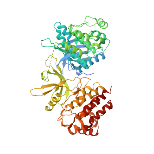
Structure of the pseudokinase-kinase domains from protein kinase TYK2 reveals a mechanism for Janus kinase (JAK) autoinhibition.

Lupardus, P.J.Ultsch, M.Wallweber, H.Bir Kohli, P.Johnson, A.R.Eigenbrot, C.

(2014) Proc Natl Acad Sci U S A 111: 8025-8030

  • DOI: https://doi.org/10.1073/pnas.1401180111
  • Primary Citation Related Structures: 
    4OLI

  • PubMed Abstract: 

    Janus kinases (JAKs) are receptor-associated multidomain tyrosine kinases that act downstream of many cytokines and interferons. JAK kinase activity is regulated by the adjacent pseudokinase domain via an unknown mechanism. Here, we report the 2.8-Å structure of the two-domain pseudokinase-kinase module from the JAK family member TYK2 in its autoinhibited form. We find that the pseudokinase and kinase interact near the kinase active site and that most reported mutations in cancer-associated JAK alleles cluster in or near this interface. Mutation of residues near the TYK2 interface that are analogous to those in cancer-associated JAK alleles, including the V617F and "exon 12" JAK2 mutations, results in increased kinase activity in vitro. These data indicate that JAK pseudokinases are autoinhibitory domains that hold the kinase domain inactive until receptor dimerization stimulates transition to an active state.


  • Organizational Affiliation
    • Departments of Structural Biology and lupardus.patrick@gene.com eigenbrot.c@gene.com.

Macromolecule Content 

  • Total Structure Weight: 73.94 kDa 
  • Atom Count: 4,396 
  • Modeled Residue Count: 539 
  • Deposited Residue Count: 645 
  • Unique protein chains: 1

Macromolecules

Find similar proteins by:|  3D Structure
Entity ID: 1
MoleculeChains  Sequence LengthOrganismDetailsImage
Non-receptor tyrosine-protein kinase TYK2645Homo sapiensMutation(s): 1 
Gene Names: TYK2
EC: 2.7.10.2
UniProt & NIH Common Fund Data Resources
Find proteins for P29597 (Homo sapiens)
Explore P29597 
Go to UniProtKB:  P29597
PHAROS:  P29597
GTEx:  ENSG00000105397 
Entity Groups
Sequence Clusters30% Identity50% Identity70% Identity90% Identity95% Identity100% Identity
UniProt GroupP29597
Sequence Annotations
Expand
Reference Sequence

Small Molecules

Ligands 1 Unique
IDChains Name / Formula / InChI Key2D Diagram3D Interactions
2TT

Query on 2TT


Download:Ideal Coordinates CCD File
B [auth A],
C [auth A]
2-chloro-N-{2-[(cyclopropylcarbonyl)amino]pyridin-4-yl}benzamide
C16 H14 Cl N3 O2
BZYFIAMMQPKLOE-UHFFFAOYSA-N

Experimental Data & Validation

Experimental Data

  • Method: X-RAY DIFFRACTION
  • Resolution: 2.80 Å
  • R-Value Free:  0.257 (Depositor), 0.264 (DCC) 
  • R-Value Work:  0.201 (Depositor), 0.211 (DCC) 
  • R-Value Observed: 0.205 (Depositor) 
Space Group: I 4
Unit Cell:
Length ( Å )Angle ( ˚ )
a = 111.015α = 90
b = 111.015β = 90
c = 123.955γ = 90
Software Package:
Software NamePurpose
BOSdata collection
PHASERphasing
BUSTERrefinement
HKL-2000data reduction
SCALEPACKdata scaling

Structure Validation

View Full Validation Report


Ligand Structure Quality Assessment 


Entry History 

Deposition Data

Revision History  (Full details and data files)

  • Version 1.0: 2014-05-28
    Type: Initial release
  • Version 1.1: 2014-07-16
    Changes: Database references
  • Version 1.2: 2023-09-20
    Changes: Data collection, Database references, Derived calculations, Refinement description