4OII | pdb_00004oii

West Nile Virus NS1 in complex with neutralizing 22NS1 antibody Fab


Experimental Data Snapshot

  • Method: X-RAY DIFFRACTION
  • Resolution: 3.00 Å
  • R-Value Free: 
    0.265 (Depositor), 0.260 (DCC) 
  • R-Value Work: 
    0.214 (Depositor), 0.211 (DCC) 
  • R-Value Observed: 
    0.216 (Depositor) 

wwPDB Validation 3D Report Full Report

Validation slider image for 4OII

This is version 1.4 of the entry. See complete history

Literature

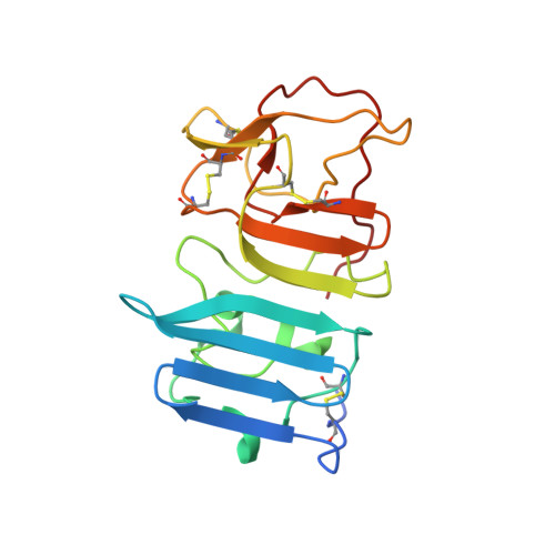
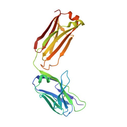
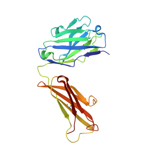
Structural basis of Flavivirus NS1 assembly and antibody recognition.

Edeling, M.A.Diamond, M.S.Fremont, D.H.

(2014) Proc Natl Acad Sci U S A 111: 4285-4290

  • DOI: https://doi.org/10.1073/pnas.1322036111
  • Primary Citation Related Structures: 
    4OIE, 4OIG, 4OII

  • PubMed Abstract: 

    The Flavivirus nonstructural protein 1 (NS1) is a conserved, membrane-associated and secreted glycoprotein with replication and immune evasion functions. Secreted NS1 is a hexameric, barrel-shaped lipoprotein that can bind back to the plasma membrane of cells. Antibodies targeting cell surface-associated NS1 can be protective in vivo in a manner dependent on Fc effector functions. We describe here the crystal structure of a C-terminal fragment (residues 172-352) of West Nile (WNV) and Dengue virus NS1 proteins at 1.85 and 2.7 Å resolution, respectively. NS1(172-352) assembles as a unique rod-shaped dimer composed of a 16-stranded β-platform flanked on one face by protruding connecting loops. We also determined the 3.0 Å resolution structure of WNV NS1(172-352) with the protective 22NS1 antibody Fab, which engages the loop-face of the rod. The head-to-head NS1(172-352) dimer we observe in crystal lattices is supported by multiangle light and small-angle X-ray scattering studies. We used the available cryo-electron microscopy reconstruction to develop a pseudoatomic model of the NS1 hexamer. The model was constructed with the NS1(172-352) dimeric rod aligned with the long axis of the barrel, and with the loop-face oriented away from the core. Difference densities suggest that the N-terminal region of NS1 forms globular lobes that mediate lateral contacts between dimers in the hexamer. Our model also suggests that the N-terminal lobe forms the surface of the central cavity where lipid binding may occur.


  • Organizational Affiliation
    • Departments of Pathology and Immunology, Medicine, Molecular Microbiology, and Biochemistry and Molecular Biophysics, Washington University in St. Louis, St. Louis, MO 63110.

Macromolecule Content 

  • Total Structure Weight: 135.47 kDa 
  • Atom Count: 9,455 
  • Modeled Residue Count: 1,214 
  • Deposited Residue Count: 1,230 
  • Unique protein chains: 3

Macromolecules

Find similar proteins by:|  3D Structure
Entity ID: 1
MoleculeChains  Sequence LengthOrganismDetailsImage
NON-STRUCTURAL PROTEIN NS1
A, B
185West Nile virusMutation(s): 0 
UniProt
Find proteins for Q9Q6P4 (West Nile virus (strain NY-99))
Explore Q9Q6P4 
Go to UniProtKB:  Q9Q6P4
Entity Groups
Sequence Clusters30% Identity50% Identity70% Identity90% Identity95% Identity100% Identity
UniProt GroupQ9Q6P4
Sequence Annotations
Expand
Reference Sequence
Find similar proteins by:|  3D Structure
Entity ID: 2
MoleculeChains  Sequence LengthOrganismDetailsImage
Light Chain of Fab fragment of 22NS1 AntibodyC [auth L],
E [auth M]
213Mus musculusMutation(s): 0 
Find similar proteins by:|  3D Structure
Entity ID: 3
MoleculeChains  Sequence LengthOrganismDetailsImage
Heavy Chain of Fab fragment of 22NS1 AntibodyD [auth H],
F [auth I]
217Mus musculusMutation(s): 0 

Experimental Data & Validation

Experimental Data

  • Method: X-RAY DIFFRACTION
  • Resolution: 3.00 Å
  • R-Value Free:  0.265 (Depositor), 0.260 (DCC) 
  • R-Value Work:  0.214 (Depositor), 0.211 (DCC) 
  • R-Value Observed: 0.216 (Depositor) 
Space Group: C 1 2 1
Unit Cell:
Length ( Å )Angle ( ˚ )
a = 215.869α = 90
b = 49.546β = 91.21
c = 130.075γ = 90
Software Package:
Software NamePurpose
SCALEPACKdata scaling
PHENIXrefinement
PDB_EXTRACTdata extraction
DENZOdata reduction
PHASERphasing

Structure Validation

View Full Validation Report


Entry History 

Deposition Data

Revision History  (Full details and data files)

  • Version 1.0: 2014-03-05
    Type: Initial release
  • Version 1.1: 2014-04-02
    Changes: Database references
  • Version 1.2: 2014-04-23
    Changes: Database references
  • Version 1.3: 2017-11-22
    Changes: Refinement description
  • Version 1.4: 2024-10-30
    Changes: Data collection, Database references, Structure summary