4OGF

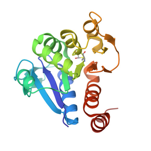
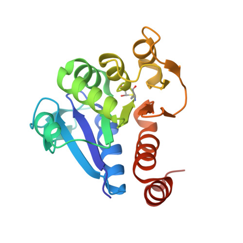
Crystal Structure of Human DJ-1 with glyoxylate as substrate analog


Experimental Data Snapshot

  • Method: X-RAY DIFFRACTION
  • Resolution: 1.60 Å
  • R-Value Free: 0.235 
  • R-Value Work: 0.212 

Starting Model: experimental
View more details

wwPDB Validation   3D Report Full Report


This is version 1.3 of the entry. See complete history


Literature

Stereospecific mechanism of DJ-1 glyoxalases inferred from their hemithioacetal-containing crystal structures.

Choi, D.Kim, J.Ha, S.Kwon, K.Kim, E.H.Lee, H.Y.Ryu, K.S.Park, C.

(2014) FEBS J 281: 5447-5462

  • DOI: https://doi.org/10.1111/febs.13085
  • Primary Citation of Related Structures:  
    4OFW, 4OGF, 4OGG

  • PubMed Abstract: 

    DJ-1 family proteins have recently been characterized as novel glyoxalases, although their cofactor-free catalytic mechanisms are not fully understood. Here, we obtained crystals of Arabidopsis thaliana DJ-1d (atDJ-1d) and Homo sapiens DJ-1 (hDJ-1) covalently bound to glyoxylate, an analog of methylglyoxal, forming a hemithioacetal that presumably mimics an intermediate structure in catalysis of methylglyoxal to lactate. The deuteration level of lactate supported the proton transfer mechanism in the enzyme reaction. Differences in the enantiomeric specificity of d/l-lactacte formation observed for the DJ-1 superfamily proteins are explained by the presence of a His residue in the active site with essential Cys and Glu residues. The model for the stereospecificity was further evaluated by a molecular modeling simulation with methylglyoxal hemithioacetal superimposed on the glyoxylate hemithioacetal. The mechanism of DJ-1 glyoxalase provides a basis for understanding the His residue-based stereospecificity. Structural data have been submitted to the Protein Data Bank under accession numbers 4OFW (structure of atDJ-1d), 4OGF (structure of hDJ-1 with glyoxylate) and 4OGG (structure of atDJ-1d with glyoxylate).


  • Organizational Affiliation

    Division of Magnetic Resonance Research, Korea Basic Science Institute, Chungcheongbuk-Do, South Korea; Department of Biological Science, Korea Advanced Institute of Science and Technology, Daejeon, South Korea.


Macromolecules
Find similar proteins by:  (by identity cutoff)  |  3D Structure
Entity ID: 1
MoleculeChains Sequence LengthOrganismDetailsImage
Protein DJ-1187Homo sapiensMutation(s): 0 
Gene Names: PARK7
EC: 4.2.1.130 (PDB Primary Data), 3.5.1 (UniProt), 3.1.2 (UniProt), 3.5.1.124 (UniProt)
UniProt & NIH Common Fund Data Resources
Find proteins for Q99497 (Homo sapiens)
Explore Q99497 
Go to UniProtKB:  Q99497
PHAROS:  Q99497
GTEx:  ENSG00000116288 
Entity Groups  
Sequence Clusters30% Identity50% Identity70% Identity90% Identity95% Identity100% Identity
UniProt GroupQ99497
Sequence Annotations
Expand
  • Reference Sequence
Small Molecules
Modified Residues  1 Unique
IDChains TypeFormula2D DiagramParent
CGV
Query on CGV
A
L-PEPTIDE LINKINGC5 H9 N O5 SCYS
Experimental Data & Validation

Experimental Data

  • Method: X-RAY DIFFRACTION
  • Resolution: 1.60 Å
  • R-Value Free: 0.235 
  • R-Value Work: 0.212 
  • Space Group: P 31 2 1
Unit Cell:
Length ( Å )Angle ( ˚ )
a = 74.692α = 90
b = 74.692β = 90
c = 74.937γ = 120
Software Package:
Software NamePurpose
SCALEPACKdata scaling
CNSrefinement
PDB_EXTRACTdata extraction
HKL-2000data reduction
Cootmodel building

Structure Validation

View Full Validation Report


Entry History 

Deposition Data

Revision History  (Full details and data files)

  • Version 1.0: 2014-10-15
    Type: Initial release
  • Version 1.1: 2014-10-22
    Changes: Database references
  • Version 1.2: 2014-12-24
    Changes: Database references
  • Version 1.3: 2023-09-20
    Changes: Data collection, Database references, Derived calculations, Refinement description