4ODR

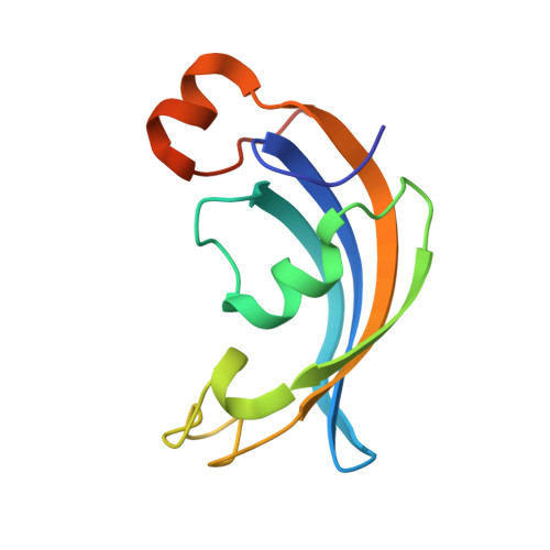
Structure of SlyD delta-IF from Thermus thermophilus in complex with FK506


Experimental Data Snapshot

  • Method: X-RAY DIFFRACTION
  • Resolution: 1.93 Å
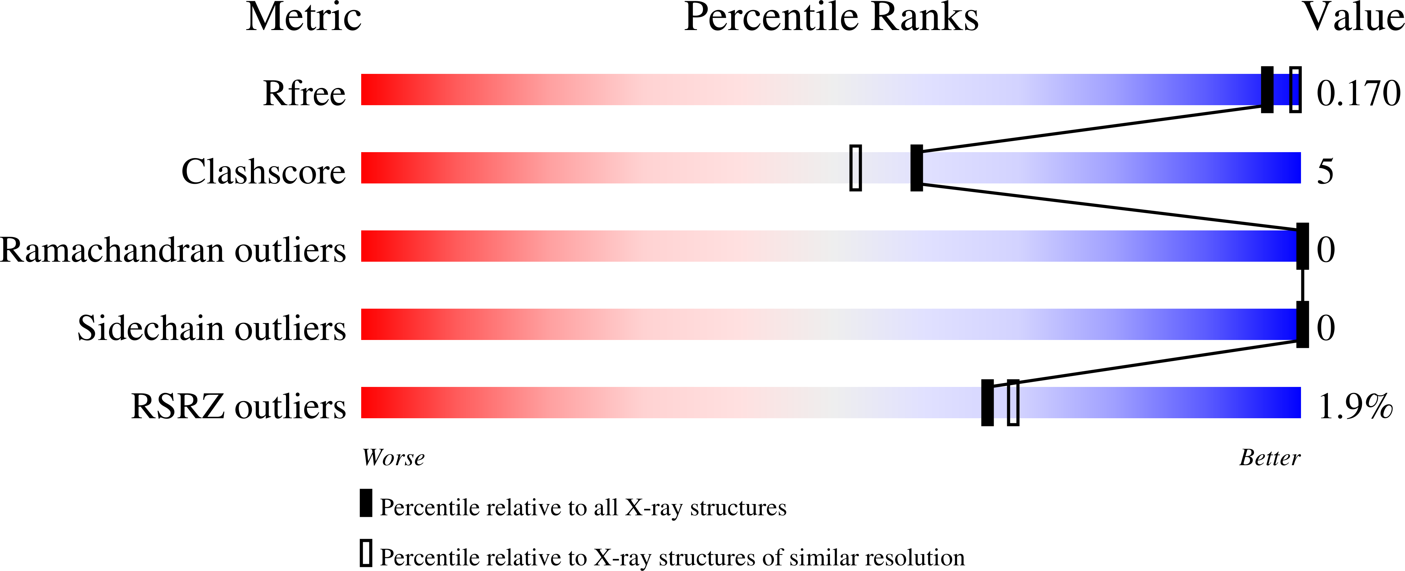
  • R-Value Free: 0.170 
  • R-Value Work: 0.153 
  • R-Value Observed: 0.153 

wwPDB Validation   3D Report Full Report


Ligand Structure Quality Assessment 


This is version 1.3 of the entry. See complete history


Literature

Molecular insights into substrate recognition and catalytic mechanism of the chaperone and FKBP peptidyl-prolyl isomerase SlyD.

Quistgaard, E.M.Weininger, U.Ural-Blimke, Y.Modig, K.Nordlund, P.Akke, M.Low, C.

(2016) BMC Biol 14: 82-82

  • DOI: https://doi.org/10.1186/s12915-016-0300-3
  • Primary Citation of Related Structures:  
    4ODK, 4ODL, 4ODM, 4ODN, 4ODO, 4ODP, 4ODQ, 4ODR

  • PubMed Abstract: 

    Peptidyl-prolyl isomerases (PPIases) catalyze cis/trans isomerization of peptidyl-prolyl bonds, which is often rate-limiting for protein folding. SlyD is a two-domain enzyme containing both a PPIase FK506-binding protein (FKBP) domain and an insert-in-flap (IF) chaperone domain. To date, the interactions of these domains with unfolded proteins have remained rather obscure, with structural information on binding to the FKBP domain being limited to complexes involving various inhibitor compounds or a chemically modified tetrapeptide. We have characterized the binding of 15-residue-long unmodified peptides to SlyD from Thermus thermophilus (TtSlyD) in terms of binding thermodynamics and enzyme kinetics through the use of isothermal titration calorimetry, nuclear magnetic resonance spectroscopy, and site-directed mutagenesis. We show that the affinities and enzymatic activity of TtSlyD towards these peptides are much higher than for the chemically modified tetrapeptides that are typically used for activity measurements on FKBPs. In addition, we present a series of crystal structures of TtSlyD with the inhibitor FK506 bound to the FKBP domain, and with 15-residue-long peptides bound to either one or both domains, which reveals that substrates bind in a highly adaptable fashion to the IF domain through β-strand augmentation, and can bind to the FKBP domain as both types VIa1 and VIb-like cis-proline β-turns. Our results furthermore provide important clues to the catalytic mechanism and support the notion of inter-domain cross talk. We found that 15-residue-long unmodified peptides can serve as better substrate mimics for the IF and FKBP domains than chemically modified tetrapeptides. We furthermore show how such peptides are recognized by each of these domains in TtSlyD, and propose a novel general model for the catalytic mechanism of FKBPs that involves C-terminal rotation around the peptidyl-prolyl bond mediated by stabilization of the twisted transition state in the hydrophobic binding site.


  • Organizational Affiliation

    Department of Medical Biochemistry and Biophysics, Karolinska Institutet, Scheeles väg 2, SE-17177, Stockholm, Sweden.


Macromolecules
Find similar proteins by:  (by identity cutoff)  |  3D Structure
Entity ID: 1
MoleculeChains Sequence LengthOrganismDetailsImage
Peptidyl-prolyl cis-trans isomerase SlyD, Peptidyl-prolyl cis-trans isomerase FKBP1A chimera
A, B
110Thermus thermophilusHomo sapiensMutation(s): 0 
Gene Names: TTHA0346FKBP1FKBP12FKBP1A
EC: 5.2.1.8
UniProt & NIH Common Fund Data Resources
Find proteins for Q5SLE7 (Thermus thermophilus (strain ATCC 27634 / DSM 579 / HB8))
Explore Q5SLE7 
Go to UniProtKB:  Q5SLE7
Find proteins for P62942 (Homo sapiens)
Explore P62942 
Go to UniProtKB:  P62942
PHAROS:  P62942
GTEx:  ENSG00000088832 
Entity Groups  
Sequence Clusters30% Identity50% Identity70% Identity90% Identity95% Identity100% Identity
UniProt GroupsQ5SLE7P62942
Sequence Annotations
Expand
  • Reference Sequence
Small Molecules
Ligands 5 Unique
IDChains Name / Formula / InChI Key2D Diagram3D Interactions
FK5
Query on FK5

Download Ideal Coordinates CCD File 
C [auth A],
M [auth B]
8-DEETHYL-8-[BUT-3-ENYL]-ASCOMYCIN
C44 H69 N O12
QJJXYPPXXYFBGM-LFZNUXCKSA-N
GOL
Query on GOL

Download Ideal Coordinates CCD File 
G [auth A]
H [auth A]
I [auth A]
J [auth A]
P [auth B]
G [auth A],
H [auth A],
I [auth A],
J [auth A],
P [auth B],
Q [auth B]
GLYCEROL
C3 H8 O3
PEDCQBHIVMGVHV-UHFFFAOYSA-N
ZN
Query on ZN

Download Ideal Coordinates CCD File 
D [auth A],
E [auth A],
N [auth B]
ZINC ION
Zn
PTFCDOFLOPIGGS-UHFFFAOYSA-N
ACT
Query on ACT

Download Ideal Coordinates CCD File 
K [auth A],
L [auth A],
R [auth B]
ACETATE ION
C2 H3 O2
QTBSBXVTEAMEQO-UHFFFAOYSA-M
CL
Query on CL

Download Ideal Coordinates CCD File 
F [auth A],
O [auth B]
CHLORIDE ION
Cl
VEXZGXHMUGYJMC-UHFFFAOYSA-M
Experimental Data & Validation

Experimental Data

  • Method: X-RAY DIFFRACTION
  • Resolution: 1.93 Å
  • R-Value Free: 0.170 
  • R-Value Work: 0.153 
  • R-Value Observed: 0.153 
  • Space Group: P 43 21 2
Unit Cell:
Length ( Å )Angle ( ˚ )
a = 72.5α = 90
b = 72.5β = 90
c = 178.96γ = 90
Software Package:
Software NamePurpose
XDSdata scaling
PHASERphasing
PHENIXrefinement
XDSdata reduction
XSCALEdata scaling

Structure Validation

View Full Validation Report


Ligand Structure Quality Assessment 


Entry History 

Deposition Data

Revision History  (Full details and data files)

  • Version 1.0: 2015-01-14
    Type: Initial release
  • Version 1.1: 2017-07-26
    Changes: Refinement description, Source and taxonomy
  • Version 1.2: 2017-10-04
    Changes: Database references
  • Version 1.3: 2024-02-28
    Changes: Data collection, Database references, Derived calculations