4O9W | pdb_00004o9w

Crystal structure of polo-like kinase(PLK1)PBD in complex with phospho peptide


Experimental Data Snapshot

  • Method: X-RAY DIFFRACTION
  • Resolution: 1.69 Å
  • R-Value Free: 
    0.240 (Depositor), 0.240 (DCC) 
  • R-Value Work: 
    0.201 (Depositor), 0.202 (DCC) 
  • R-Value Observed: 
    0.203 (Depositor) 

wwPDB Validation 3D Report Full Report

Validation slider image for 4O9W

This is version 1.3 of the entry. See complete history

Literature

The condensin component NCAPG2 regulates microtubule-kinetochore attachment through recruitment of Polo-like kinase 1 to kinetochores.

Kim, J.H.Shim, J.Ji, M.J.Jung, Y.Bong, S.M.Jang, Y.J.Yoon, E.K.Lee, S.J.Kim, K.G.Kim, Y.H.Lee, C.Lee, B.I.Kim, K.T.

(2014) Nat Commun 5: 4588-4588

  • DOI: https://doi.org/10.1038/ncomms5588
  • Primary Citation Related Structures: 
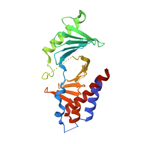
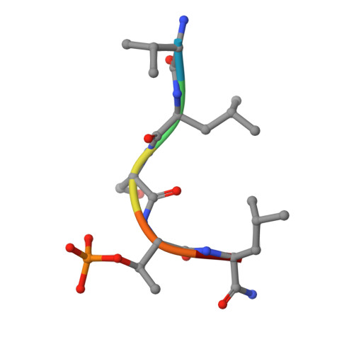
    4O9W

  • PubMed Abstract: 

    The early event of microtubule-kinetochore attachment is a critical stage for precise chromosome segregation. Here we report that NCAPG2, which is a component of the condensin II complex, mediates chromosome segregation through microtubule-kinetochore attachment by recruiting PLK1 to prometaphase kinetochores. NCAPG2 colocalizes with PLK1 at prometaphase kinetochores and directly interacts with the polo-box domain (PBD) of PLK1 via its highly conserved C-terminal region. In both humans and Caenorhabditis elegans, when NCAPG2 is depleted, the attachment of the spindle to the kinetochore is loosened and misoriented. This is caused by the disruption of PLK1 localization to the kinetochore and by the decreased phosphorylation of its kinetochore substrate, BubR1. In addition, the crystal structure of the PBD of PLK1, in complex with the C-terminal region of NCAPG2, (1007)VLS-pT-L(1011), exhibits structural conservation of PBD-phosphopeptides, suggesting that the regulation of NCAPG2 function is phosphorylation-dependent. These findings suggest that NCAPG2 plays an important role in regulating proper chromosome segregation through a functional interaction with PLK1 during mitosis.


  • Organizational Affiliation
    • 1] Research Institute, National Cancer Center, Goyang, Gyeonggi 410-769, Republic of Korea [2].

Macromolecule Content 

  • Total Structure Weight: 26.36 kDa 
  • Atom Count: 1,825 
  • Modeled Residue Count: 217 
  • Deposited Residue Count: 228 
  • Unique protein chains: 2

Macromolecules

Find similar proteins by:|  3D Structure
Entity ID: 1
MoleculeChains  Sequence LengthOrganismDetailsImage
Serine/threonine-protein kinase PLK1222Homo sapiensMutation(s): 0 
Gene Names: PLK1PLK
EC: 2.7.11.21
UniProt & NIH Common Fund Data Resources
Find proteins for P53350 (Homo sapiens)
Explore P53350 
Go to UniProtKB:  P53350
PHAROS:  P53350
GTEx:  ENSG00000166851 
Entity Groups
Sequence Clusters30% Identity50% Identity70% Identity90% Identity95% Identity100% Identity
UniProt GroupP53350
Sequence Annotations
Expand
Reference Sequence
Find similar proteins by:  Sequence   |   3D Structure  
Entity ID: 2
MoleculeChains  Sequence LengthOrganismDetailsImage
phospho peptide VAL-LEU-SER-TPO-LEU-NH26Homo sapiensMutation(s): 0 

Small Molecules

Modified Residues  1 Unique
IDChains TypeFormula2D DiagramParent
TPO
Query on TPO
B
L-PEPTIDE LINKINGC4 H10 N O6 PTHR

Experimental Data & Validation

Experimental Data

  • Method: X-RAY DIFFRACTION
  • Resolution: 1.69 Å
  • R-Value Free:  0.240 (Depositor), 0.240 (DCC) 
  • R-Value Work:  0.201 (Depositor), 0.202 (DCC) 
  • R-Value Observed: 0.203 (Depositor) 
Space Group: P 1 21 1
Unit Cell:
Length ( Å )Angle ( ˚ )
a = 35.649α = 90
b = 51.858β = 100.69
c = 57.654γ = 90
Software Package:
Software NamePurpose
HKL-2000data collection
PHASERphasing
PHENIXrefinement
HKL-2000data reduction
HKL-2000data scaling

Structure Validation

View Full Validation Report


Entry History 

Deposition Data

Revision History  (Full details and data files)

  • Version 1.0: 2014-08-13
    Type: Initial release
  • Version 1.1: 2014-11-26
    Changes: Other
  • Version 1.2: 2019-12-18
    Changes: Advisory, Database references, Derived calculations
  • Version 1.3: 2024-10-09
    Changes: Advisory, Data collection, Database references, Structure summary