4O9A | pdb_00004o9a

Crystal structure of Beta-ketothiolase (PhaA) from Ralstonia eutropha H16


Experimental Data Snapshot

  • Method: X-RAY DIFFRACTION
  • Resolution: 1.52 Å
  • R-Value Free: 
    0.254 (Depositor), 0.257 (DCC) 
  • R-Value Work: 
    0.218 (Depositor), 0.222 (DCC) 
  • R-Value Observed: 
    0.219 (Depositor) 

Starting Model: experimental
View more details

wwPDB Validation 3D Report Full Report

Validation slider image for 4O9A

This is version 1.2 of the entry. See complete history

Literature

Crystal structure and biochemical characterization of PhaA from Ralstonia eutropha, a polyhydroxyalkanoate-producing bacterium.

Kim, E.J.Kim, K.J.

(2014) Biochem Biophys Res Commun 452: 124-129

  • DOI: https://doi.org/10.1016/j.bbrc.2014.08.074
  • Primary Citation Related Structures: 
    4O99, 4O9A, 4O9C

  • PubMed Abstract: 

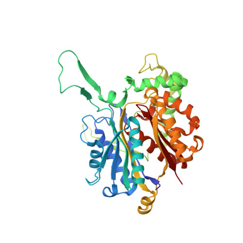
    PhaA from Ralstonia eutropha (RePhaA) is the first enzyme in the polyhydroxyalbutyrate (PHB) biosynthetic pathway and catalyzes the condensation of two molecules of acetyl-CoA to acetoacetyl-CoA. To investigate the molecular mechanism underlying PHB biosynthesis, we determined the crystal structures of the RePhaA protein in apo- and CoA-bound forms. The RePhaA structure adopts the type II biosynthetic thiolase fold forming a tetramer by means of dimerization of two dimers. The crystal structure of RePhaA in complex with CoA revealed that the enzyme contained a unique Phe219 residue, resulting that the ADP moiety binds in somewhat different position compared with that bound in other thiolase enzymes. Our study provides structural insight into the substrate specificity of RePhaA. Results indicate the presence of a small pocket near the Cys88 covalent catalytic residue leading to the possibility of the enzyme to accommodate acetyl-CoA as a sole substrate instead of larger acyl-CoA molecules such as propionyl-CoA. Furthermore, the roles of key residues involved in substrate binding and enzyme catalysis were confirmed by site-directed mutagenesis.


  • Organizational Affiliation
    • School of Life Sciences, KNU Creative BioResearch Group, Kyungpook National University, Daehak-ro 80, Buk-ku, Daegu 702-701, Republic of Korea.

Macromolecule Content 

  • Total Structure Weight: 165.1 kDa 
  • Atom Count: 11,616 
  • Modeled Residue Count: 1,568 
  • Deposited Residue Count: 1,592 
  • Unique protein chains: 1

Macromolecules

Find similar proteins by:|  3D Structure
Entity ID: 1
MoleculeChains  Sequence LengthOrganismDetailsImage
Acetyl-CoA acetyltransferase
A, B, C, D
398Cupriavidus necator H16Mutation(s): 1 
Gene Names: H16_A1438phaAphbA
EC: 2.3.1.9
UniProt
Find proteins for P14611 (Cupriavidus necator (strain ATCC 17699 / DSM 428 / KCTC 22496 / NCIMB 10442 / H16 / Stanier 337))
Explore P14611 
Go to UniProtKB:  P14611
Entity Groups
Sequence Clusters30% Identity50% Identity70% Identity90% Identity95% Identity100% Identity
UniProt GroupP14611
Sequence Annotations
Expand
Reference Sequence

Experimental Data & Validation

Experimental Data

  • Method: X-RAY DIFFRACTION
  • Resolution: 1.52 Å
  • R-Value Free:  0.254 (Depositor), 0.257 (DCC) 
  • R-Value Work:  0.218 (Depositor), 0.222 (DCC) 
  • R-Value Observed: 0.219 (Depositor) 
Space Group: P 1 21 1
Unit Cell:
Length ( Å )Angle ( ˚ )
a = 68.271α = 90
b = 104.705β = 106.12
c = 106.907γ = 90
Software Package:
Software NamePurpose
REFMACrefinement
PDB_EXTRACTdata extraction
DENZOdata reduction
SCALEPACKdata scaling

Structure Validation

View Full Validation Report


Entry History 

Deposition Data

Revision History  (Full details and data files)

  • Version 1.0: 2015-02-04
    Type: Initial release
  • Version 1.1: 2017-11-22
    Changes: Refinement description
  • Version 1.2: 2023-11-08
    Changes: Data collection, Database references, Refinement description