4O8X | pdb_00004o8x

Zinc-bound Rpn11 in complex with Rpn8


Experimental Data Snapshot

  • Method: X-RAY DIFFRACTION
  • Resolution: 1.99 Å
  • R-Value Free: 
    0.195 (Depositor), 0.196 (DCC) 
  • R-Value Work: 
    0.161 (Depositor), 0.166 (DCC) 
  • R-Value Observed: 
    0.165 (Depositor) 

Starting Model: experimental
View more details

wwPDB Validation 3D Report Full Report

Validation slider image for 4O8X

This is version 1.4 of the entry. See complete history

Literature

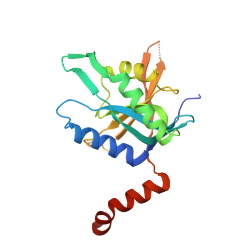
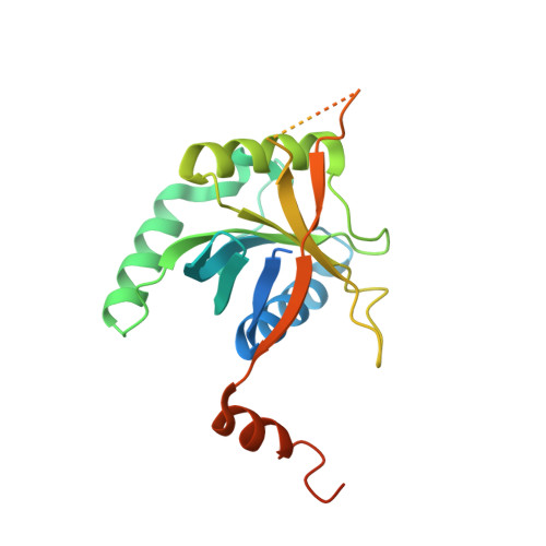
Structure of the Rpn11-Rpn8 dimer reveals mechanisms of substrate deubiquitination during proteasomal degradation.

Worden, E.J.Padovani, C.Martin, A.

(2014) Nat Struct Mol Biol 21: 220-227

  • DOI: https://doi.org/10.1038/nsmb.2771
  • Primary Citation Related Structures: 
    4O8X, 4O8Y

  • PubMed Abstract: 

    Polyubiquitin chains target protein substrates to the 26S proteasome, where they are removed by the deubiquitinase Rpn11 to allow efficient substrate degradation. Despite Rpn11's essential function during substrate processing, its detailed structural and biochemical characterization has been hindered by difficulties in purifying the isolated enzyme. Here we report the 2.0-Å crystal structures of Zn(2+)-free and Zn(2+)-bound Saccharomyces cerevisiae Rpn11 in an MPN-domain heterodimer with Rpn8. The Rpn11-Rpn8 interaction occurs via two distinct interfaces that may be conserved in related MPN-domain complexes. Our structural and mutational studies reveal that Rpn11 lacks a conserved surface to bind the ubiquitin Ile44 patch, does not interact with the moiety on the proximal side of the scissile isopeptide bond and exhibits no linkage specificity for ubiquitin cleavage. These findings explain how Rpn11 functions as a promiscuous deubiquitinase for cotranslocational substrate deubiquitination during proteasomal degradation.


  • Organizational Affiliation
    • Department of Molecular and Cell Biology, University of California, Berkeley, Berkeley, California, USA.

Macromolecule Content 

  • Total Structure Weight: 49.92 kDa 
  • Atom Count: 3,082 
  • Modeled Residue Count: 340 
  • Deposited Residue Count: 425 
  • Unique protein chains: 2

Macromolecules

Find similar proteins by:|  3D Structure
Entity ID: 1
MoleculeChains  Sequence LengthOrganismDetailsImage
26S proteasome regulatory subunit RPN8185Saccharomyces cerevisiae S288CMutation(s): 0 
Gene Names: RPN8YOR261CO5360
UniProt
Find proteins for Q08723 (Saccharomyces cerevisiae (strain ATCC 204508 / S288c))
Explore Q08723 
Go to UniProtKB:  Q08723
Entity Groups
Sequence Clusters30% Identity50% Identity70% Identity90% Identity95% Identity100% Identity
UniProt GroupQ08723
Sequence Annotations
Expand
Reference Sequence
Find similar proteins by:|  3D Structure
Entity ID: 2
MoleculeChains  Sequence LengthOrganismDetailsImage
26S proteasome regulatory subunit RPN11240Saccharomyces cerevisiae S288CMutation(s): 0 
Gene Names: RPN11MPR1YFR004W
EC: 3.4.19.12
UniProt
Find proteins for P43588 (Saccharomyces cerevisiae (strain ATCC 204508 / S288c))
Explore P43588 
Go to UniProtKB:  P43588
Entity Groups
Sequence Clusters30% Identity50% Identity70% Identity90% Identity95% Identity100% Identity
UniProt GroupP43588
Sequence Annotations
Expand
Reference Sequence

Small Molecules

Ligands 2 Unique
IDChains Name / Formula / InChI Key2D Diagram3D Interactions
ZN

Query on ZN


Download:Ideal Coordinates CCD File
HA [auth B]ZINC ION
Zn
PTFCDOFLOPIGGS-UHFFFAOYSA-N
EDO

Query on EDO


Download:Ideal Coordinates CCD File
AA [auth B]
BA [auth B]
C [auth A]
CA [auth B]
D [auth A]
AA [auth B],
BA [auth B],
C [auth A],
CA [auth B],
D [auth A],
DA [auth B],
E [auth A],
EA [auth B],
F [auth A],
FA [auth B],
G [auth A],
GA [auth B],
H [auth A],
I [auth A],
J [auth A],
K [auth A],
L [auth A],
M [auth A],
N [auth A],
O [auth A],
P [auth A],
Q [auth A],
R [auth A],
S [auth A],
T [auth B],
U [auth B],
V [auth B],
W [auth B],
X [auth B],
Y [auth B],
Z [auth B]
1,2-ETHANEDIOL
C2 H6 O2
LYCAIKOWRPUZTN-UHFFFAOYSA-N

Experimental Data & Validation

Experimental Data

  • Method: X-RAY DIFFRACTION
  • Resolution: 1.99 Å
  • R-Value Free:  0.195 (Depositor), 0.196 (DCC) 
  • R-Value Work:  0.161 (Depositor), 0.166 (DCC) 
  • R-Value Observed: 0.165 (Depositor) 
Space Group: P 43 21 2
Unit Cell:
Length ( Å )Angle ( ˚ )
a = 70.296α = 90
b = 70.296β = 90
c = 198.912γ = 90
Software Package:
Software NamePurpose
DENZOdata reduction
SCALEPACKdata scaling
PHASERphasing
PHENIXrefinement
PDB_EXTRACTdata extraction
Blu-Icedata collection

Structure Validation

View Full Validation Report


Entry History 

Deposition Data

Revision History  (Full details and data files)

  • Version 1.0: 2014-01-22
    Type: Initial release
  • Version 1.1: 2014-02-26
    Changes: Database references
  • Version 1.2: 2014-03-19
    Changes: Database references
  • Version 1.3: 2017-11-22
    Changes: Refinement description
  • Version 1.4: 2023-09-20
    Changes: Data collection, Database references, Derived calculations, Refinement description