4O4D | pdb_00004o4d

Crystal Structure of an Inositol hexakisphosphate kinase EhIP6KA in complexed with ATP and Ins(1,4,5)P3


Experimental Data Snapshot

  • Method: X-RAY DIFFRACTION
  • Resolution: 2.10 Å
  • R-Value Free: 
    0.227 (Depositor), 0.209 (DCC) 
  • R-Value Work: 
    0.173 (Depositor) 
  • R-Value Observed: 
    0.176 (Depositor) 

Starting Model: experimental
View more details

wwPDB Validation 3D Report Full Report

Validation slider image for 4O4D

Ligand Structure Quality Assessment 


This is version 1.2 of the entry. See complete history

Literature

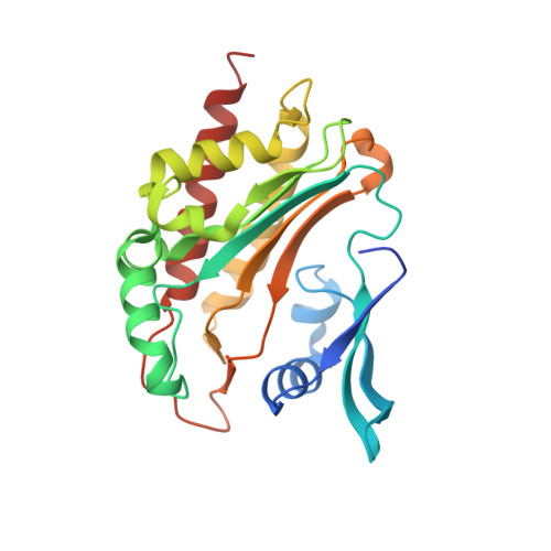
IP6K structure and the molecular determinants of catalytic specificity in an inositol phosphate kinase family.

Wang, H.DeRose, E.F.London, R.E.Shears, S.B.

(2014) Nat Commun 5: 4178-4178

  • DOI: https://doi.org/10.1038/ncomms5178
  • Primary Citation Related Structures: 
    4O4B, 4O4C, 4O4D, 4O4E, 4O4F

  • PubMed Abstract: 

    Inositol trisphosphate kinases (IP3Ks) and inositol hexakisphosphate kinases (IP6Ks) each regulate specialized signalling activities by phosphorylating either InsP3 or InsP6 respectively. The molecular basis for these different kinase activities can be illuminated by a structural description of IP6K. Here we describe the crystal structure of an Entamoeba histolytica hybrid IP6K/IP3K, an enzymatic parallel to a 'living fossil'. Through molecular modelling and mutagenesis, we extrapolated our findings to human IP6K2, which retains vestigial IP3K activity. Two structural elements, an α-helical pair and a rare, two-turn 310 helix, together forge a substrate-binding pocket with an open clamshell geometry. InsP6 forms substantial contacts with both structural elements. Relative to InsP6, enzyme-bound InsP3 rotates 55° closer to the α-helices, which provide most of the protein's interactions with InsP3. These data reveal the molecular determinants of IP6K activity, and suggest an unusual evolutionary trajectory for a primordial kinase that could have favored efficient bifunctionality, before propagation of separate IP3Ks and IP6Ks.


  • Organizational Affiliation
    • Inositol Signaling Group, National Institute of Environmental Health Sciences, National Institutes of Health, Research Triangle Park, North Carolina, NC 27709, USA.

Macromolecule Content 

  • Total Structure Weight: 30.49 kDa 
  • Atom Count: 2,335 
  • Modeled Residue Count: 240 
  • Deposited Residue Count: 248 
  • Unique protein chains: 1

Macromolecules

Find similar proteins by:|  3D Structure
Entity ID: 1
MoleculeChains  Sequence LengthOrganismDetailsImage
Inositol hexakisphosphate kinase248Entamoeba histolytica HM-1:IMSS-AMutation(s): 0 
Gene Names: EHI7A_103520
EC: 2.7.4.21 (PDB Primary Data), 2.7 (UniProt)
UniProt
Find proteins for N9UNA8 (Entamoeba histolytica HM-1:IMSS-A)
Explore N9UNA8 
Go to UniProtKB:  N9UNA8
Entity Groups
Sequence Clusters30% Identity50% Identity70% Identity90% Identity95% Identity100% Identity
UniProt GroupN9UNA8
Sequence Annotations
Expand
Reference Sequence

Small Molecules

Ligands 5 Unique
IDChains Name / Formula / InChI Key2D Diagram3D Interactions
ATP

Query on ATP


Download:Ideal Coordinates CCD File
E [auth A]ADENOSINE-5'-TRIPHOSPHATE
C10 H16 N5 O13 P3
ZKHQWZAMYRWXGA-KQYNXXCUSA-N
I3P

Query on I3P


Download:Ideal Coordinates CCD File
D [auth A]D-MYO-INOSITOL-1,4,5-TRIPHOSPHATE
C6 H15 O15 P3
MMWCIQZXVOZEGG-XJTPDSDZSA-N
PO4

Query on PO4


Download:Ideal Coordinates CCD File
F [auth A]PHOSPHATE ION
O4 P
NBIIXXVUZAFLBC-UHFFFAOYSA-K
ACT

Query on ACT


Download:Ideal Coordinates CCD File
G [auth A]ACETATE ION
C2 H3 O2
QTBSBXVTEAMEQO-UHFFFAOYSA-M
MG

Query on MG


Download:Ideal Coordinates CCD File
B [auth A],
C [auth A]
MAGNESIUM ION
Mg
JLVVSXFLKOJNIY-UHFFFAOYSA-N

Experimental Data & Validation

Experimental Data

  • Method: X-RAY DIFFRACTION
  • Resolution: 2.10 Å
  • R-Value Free:  0.227 (Depositor), 0.209 (DCC) 
  • R-Value Work:  0.173 (Depositor) 
  • R-Value Observed: 0.176 (Depositor) 
Space Group: I 41 2 2
Unit Cell:
Length ( Å )Angle ( ˚ )
a = 102.738α = 90
b = 102.738β = 90
c = 111.737γ = 90
Software Package:
Software NamePurpose
HKL-2000data collection
CCP4model building
REFMACrefinement
HKL-2000data reduction
HKL-2000data scaling
CCP4phasing

Structure Validation

View Full Validation Report


Ligand Structure Quality Assessment 


Entry History 

Deposition Data

Revision History  (Full details and data files)

  • Version 1.0: 2014-06-18
    Type: Initial release
  • Version 1.1: 2014-07-09
    Changes: Database references
  • Version 1.2: 2023-09-20
    Changes: Data collection, Database references, Derived calculations, Refinement description