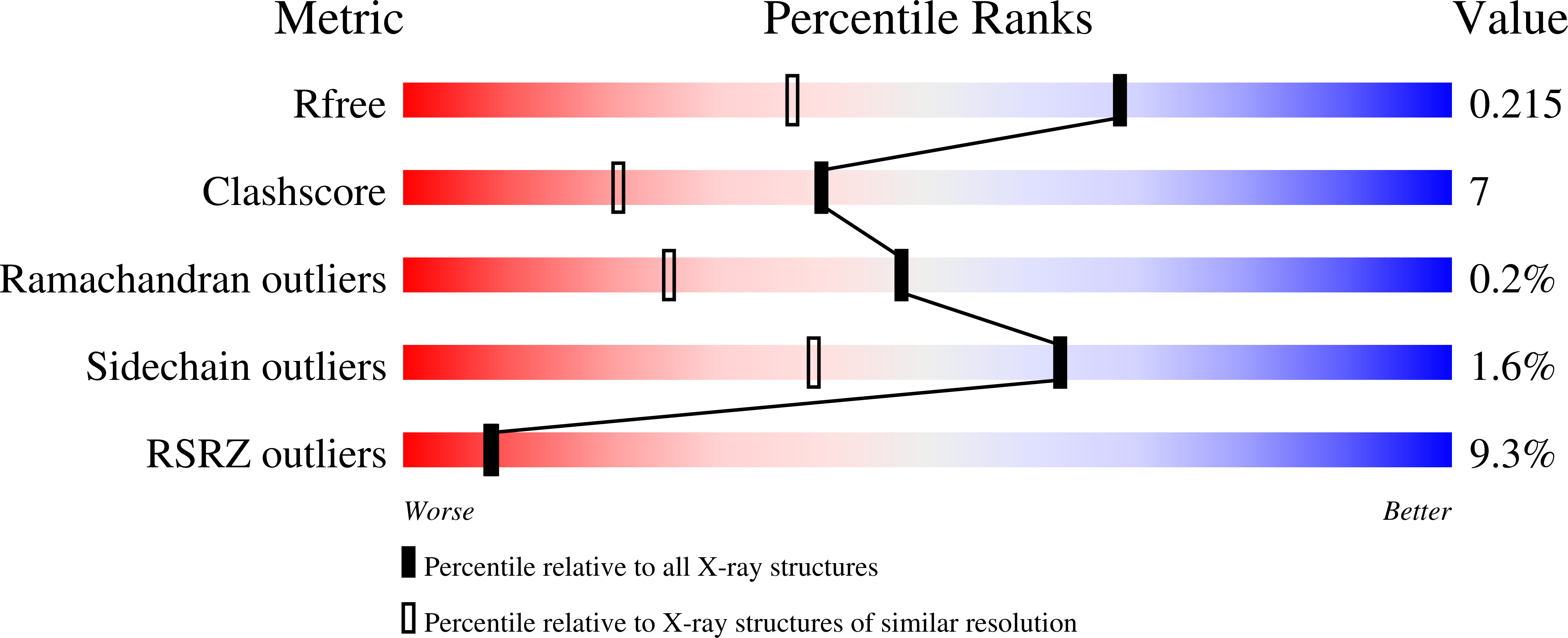
4O3N

Crystal structure of human dna polymerase eta in ternary complex with native dna and incoming nucleotide (dcp)


Experimental Data Snapshot

  • Method: X-RAY DIFFRACTION
  • Resolution: 1.58 Å
  • R-Value Free: 
    0.215 (Depositor), 0.220 (DCC) 
  • R-Value Work: 
    0.170 (Depositor), 0.170 (DCC) 
  • R-Value Observed: 
    0.172 (Depositor) 

Starting Model: experimental
View more details

wwPDB Validation   3D Report Full Report


Ligand Structure Quality Assessment 

Created with Raphaël 2.3.0Worse 01 BetterLigand structure goodness of fit to experimental dataBest fitted 0KXClick on this verticalbar to view details

This is version 1.2 of the entry. See complete history


Literature

Kinetics, Structure, and Mechanism of 8-Oxo-7,8-dihydro-2'-deoxyguanosine Bypass by Human DNA Polymerase eta

Patra, A.Nagy, L.D.Zhang, Q.Su, Y.Muller, L.Guengerich, F.P.Egli, M.

(2014) J Biol Chem 289: 16867-16882

  • DOI: https://doi.org/10.1074/jbc.M114.551820
  • Primary Citation of Related Structures:  
    4O3N, 4O3O, 4O3P, 4O3Q, 4O3R, 4O3S

  • PubMed Abstract: 

    DNA damage incurred by a multitude of endogenous and exogenous factors constitutes an inevitable challenge for the replication machinery. Cells rely on various mechanisms to either remove lesions or bypass them in a more or less error-prone fashion. The latter pathway involves the Y-family polymerases that catalyze trans-lesion synthesis across sites of damaged DNA. 7,8-Dihydro-8-oxo-2'-deoxyguanosine (8-oxoG) is a major lesion that is a consequence of oxidative stress and is associated with cancer, aging, hepatitis, and infertility. We have used steady-state and transient-state kinetics in conjunction with mass spectrometry to analyze in vitro bypass of 8-oxoG by human DNA polymerase η (hpol η). Unlike the high fidelity polymerases that show preferential insertion of A opposite 8-oxoG, hpol η is capable of bypassing 8-oxoG in a mostly error-free fashion, thus preventing GC→AT transversion mutations. Crystal structures of ternary hpol η-DNA complexes and incoming dCTP, dATP, or dGTP opposite 8-oxoG reveal that an arginine from the finger domain assumes a key role in avoiding formation of the nascent 8-oxoG:A pair. That hpol η discriminates against dATP exclusively at the insertion stage is confirmed by structures of ternary complexes that allow visualization of the extension step. These structures with G:dCTP following either 8-oxoG:C or 8-oxoG:A pairs exhibit virtually identical active site conformations. Our combined data provide a detailed understanding of hpol η bypass of the most common oxidative DNA lesion.


  • Organizational Affiliation

    From the Department of Biochemistry and Center in Molecular Toxicology, Vanderbilt University School of Medicine, Nashville, Tennessee 37232 and.


Macromolecules

Find similar proteins by:  (by identity cutoff)  |  3D Structure
Entity ID: 1
MoleculeChains Sequence LengthOrganismDetailsImage
DNA polymerase eta435Homo sapiensMutation(s): 0 
Gene Names: POLHRAD30RAD30AXPV
EC: 2.7.7.7
UniProt & NIH Common Fund Data Resources
Find proteins for Q9Y253 (Homo sapiens)
Explore Q9Y253 
Go to UniProtKB:  Q9Y253
PHAROS:  Q9Y253
GTEx:  ENSG00000170734 
Entity Groups  
Sequence Clusters30% Identity50% Identity70% Identity90% Identity95% Identity100% Identity
UniProt GroupQ9Y253
Sequence Annotations
Expand
  • Reference Sequence
Experimental Data & Validation

Experimental Data

  • Method: X-RAY DIFFRACTION
  • Resolution: 1.58 Å
  • R-Value Free:  0.215 (Depositor), 0.220 (DCC) 
  • R-Value Work:  0.170 (Depositor), 0.170 (DCC) 
  • R-Value Observed: 0.172 (Depositor) 
Space Group: P 61
Unit Cell:
Length ( Å )Angle ( ˚ )
a = 98.662α = 90
b = 98.662β = 90
c = 81.787γ = 120
Software Package:
Software NamePurpose
MD2data collection
PHASERphasing
PHENIXrefinement
HKL-2000data reduction
HKL-2000data scaling

Structure Validation

View Full Validation Report


Ligand Structure Quality Assessment 

Created with Raphaël 2.3.0Worse 01 BetterLigand structure goodness of fit to experimental dataBest fitted 0KXClick on this verticalbar to view details

Entry History 

Deposition Data

Revision History  (Full details and data files)

  • Version 1.0: 2014-04-30
    Type: Initial release
  • Version 1.1: 2014-07-02
    Changes: Database references
  • Version 1.2: 2023-09-20
    Changes: Data collection, Database references, Derived calculations, Refinement description