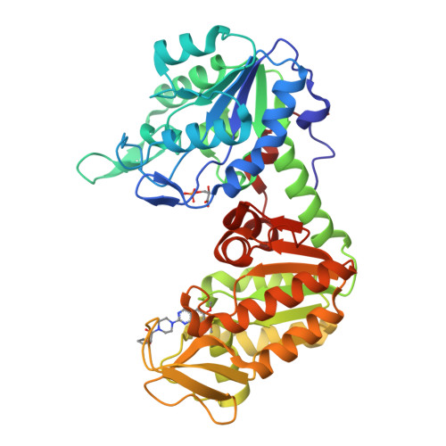
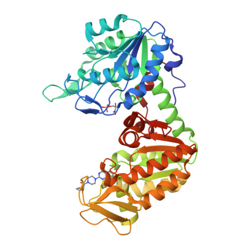
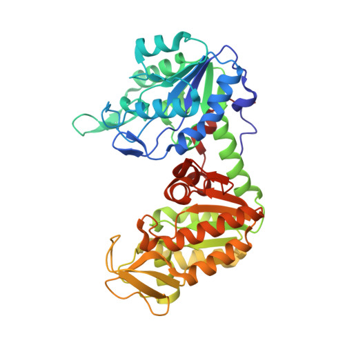
4O33

Crystal Structure of human PGK1 3PG and terazosin(TZN) ternary complex


Experimental Data Snapshot

  • Method: X-RAY DIFFRACTION
  • Resolution: 2.10 Å
  • R-Value Free: 0.221 
  • R-Value Work: 0.193 
  • R-Value Observed: 0.196 

Starting Model: experimental
View more details

wwPDB Validation   3D Report Full Report


Ligand Structure Quality Assessment 


This is version 1.3 of the entry. See complete history


Literature

Terazosin activates Pgk1 and Hsp90 to promote stress resistance.

Chen, X.Zhao, C.Li, X.Wang, T.Li, Y.Cao, C.Ding, Y.Dong, M.Finci, L.Wang, J.H.Li, X.Liu, L.

(2015) Nat Chem Biol 11: 19-25

  • DOI: https://doi.org/10.1038/nchembio.1657
  • Primary Citation of Related Structures:  
    4O33, 4O3F

  • PubMed Abstract: 

    Drugs that can protect against organ damage are urgently needed, especially for diseases such as sepsis and brain stroke. We discovered that terazosin (TZ), a widely marketed α1-adrenergic receptor antagonist, alleviated organ damage and improved survival in rodent models of stroke and sepsis. Through combined studies of enzymology and X-ray crystallography, we discovered that TZ binds a new target, phosphoglycerate kinase 1 (Pgk1), and activates its enzymatic activity, probably through 2,4-diamino-6,7-dimethoxyisoquinoline's ability to promote ATP release from Pgk1. Mechanistically, the ATP generated from Pgk1 may enhance the chaperone activity of Hsp90, an ATPase known to associate with Pgk1. Upon activation, Hsp90 promotes multistress resistance. Our studies demonstrate that TZ has a new protein target, Pgk1, and reveal its corresponding biological effect. As a clinical drug, TZ may be quickly translated into treatments for diseases including stroke and sepsis.


  • Organizational Affiliation

    1] State Key Laboratory of Biomembrane and Membrane Biotechnology, School of Life Sciences, Peking University, Beijing, China. [2] Beijing Institute for Brain Disorder and Beijing Tiantan Hospital, Capital Medical University, Beijing, China.


Macromolecules
Find similar proteins by:  (by identity cutoff)  |  3D Structure
Entity ID: 1
MoleculeChains Sequence LengthOrganismDetailsImage
Phosphoglycerate kinase 1417Homo sapiensMutation(s): 0 
Gene Names: PGK1PGKAMIG10OK/SW-cl.110
EC: 2.7.2.3
UniProt & NIH Common Fund Data Resources
Find proteins for P00558 (Homo sapiens)
Explore P00558 
Go to UniProtKB:  P00558
PHAROS:  P00558
GTEx:  ENSG00000102144 
Entity Groups  
Sequence Clusters30% Identity50% Identity70% Identity90% Identity95% Identity100% Identity
UniProt GroupP00558
Sequence Annotations
Expand
  • Reference Sequence
Small Molecules
Ligands 2 Unique
IDChains Name / Formula / InChI Key2D Diagram3D Interactions
TZN
Query on TZN

Download Ideal Coordinates CCD File 
B [auth A][4-(4-amino-6,7-dimethoxyquinazolin-2-yl)piperazin-1-yl][(2R)-tetrahydrofuran-2-yl]methanone
C19 H25 N5 O4
VCKUSRYTPJJLNI-CQSZACIVSA-N
3PG
Query on 3PG

Download Ideal Coordinates CCD File 
C [auth A]3-PHOSPHOGLYCERIC ACID
C3 H7 O7 P
OSJPPGNTCRNQQC-UWTATZPHSA-N
Experimental Data & Validation

Experimental Data

  • Method: X-RAY DIFFRACTION
  • Resolution: 2.10 Å
  • R-Value Free: 0.221 
  • R-Value Work: 0.193 
  • R-Value Observed: 0.196 
  • Space Group: P 21 21 2
Unit Cell:
Length ( Å )Angle ( ˚ )
a = 61.817α = 90
b = 73.754β = 90
c = 93.466γ = 90
Software Package:
Software NamePurpose
HKL-2000data collection
CCP4model building
PHENIXrefinement
HKL-2000data reduction
HKL-2000data scaling
CCP4phasing

Structure Validation

View Full Validation Report


Ligand Structure Quality Assessment 


Entry History 

Deposition Data

Revision History  (Full details and data files)

  • Version 1.0: 2014-10-29
    Type: Initial release
  • Version 1.1: 2014-11-26
    Changes: Database references
  • Version 1.2: 2014-12-31
    Changes: Database references
  • Version 1.3: 2023-09-20
    Changes: Data collection, Database references, Derived calculations, Refinement description