4O1P | pdb_00004o1p

Crystal Structure of RNase L in complex with 2-5A and AMP-PNP


Experimental Data Snapshot

  • Method: X-RAY DIFFRACTION
  • Resolution: 2.50 Å
  • R-Value Free: 
    0.231 (Depositor), 0.232 (DCC) 
  • R-Value Work: 
    0.196 (Depositor), 0.197 (DCC) 
  • R-Value Observed: 
    0.198 (Depositor) 

Starting Model: experimental
View more details

wwPDB Validation 3D Report Full Report

Validation slider image for 4O1P

Ligand Structure Quality Assessment 


This is version 1.2 of the entry. See complete history

Literature

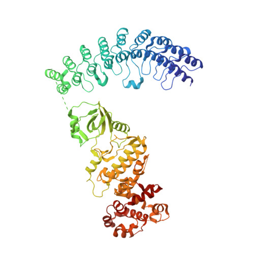
Dimeric structure of pseudokinase RNase L bound to 2-5A reveals a basis for interferon-induced antiviral activity.

Huang, H.Zeqiraj, E.Dong, B.Jha, B.K.Duffy, N.M.Orlicky, S.Thevakumaran, N.Talukdar, M.Pillon, M.C.Ceccarelli, D.F.Wan, L.C.Juang, Y.C.Mao, D.Y.Gaughan, C.Brinton, M.A.Perelygin, A.A.Kourinov, I.Guarne, A.Silverman, R.H.Sicheri, F.

(2014) Mol Cell 53: 221-234

  • DOI: https://doi.org/10.1016/j.molcel.2013.12.025
  • Primary Citation Related Structures: 
    4O1O, 4O1P

  • PubMed Abstract: 

    RNase L is an ankyrin repeat domain-containing dual endoribonuclease-pseudokinase that is activated by unusual 2,'5'-oligoadenylate (2-5A) second messengers and which impedes viral infections in higher vertebrates. Despite its importance in interferon-regulated antiviral innate immunity, relatively little is known about its precise mechanism of action. Here we present a functional characterization of 2.5 Å and 3.25 Å X-ray crystal and small-angle X-ray scattering structures of RNase L bound to a natural 2-5A activator with and without ADP or the nonhydrolysable ATP mimetic AMP-PNP. These studies reveal how recognition of 2-5A through interactions with the ankyrin repeat domain and the pseudokinase domain, together with nucleotide binding, imposes a rigid intertwined dimer configuration that is essential for RNase catalytic and antiviral functions. The involvement of the pseudokinase domain of RNase L in 2-5A sensing, nucleotide binding, dimerization, and ribonuclease functions highlights the evolutionary adaptability of the eukaryotic protein kinase fold.


  • Organizational Affiliation
    • Program in Systems Biology, Lunenfeld-Tanenbaum Research Institute, Mount Sinai Hospital, 600 University Avenue, Toronto, ON M5G 1X5, Canada; Department of Molecular Genetics, University of Toronto, Toronto, ON M5S 1A8, Canada.

Macromolecule Content 

  • Total Structure Weight: 332.19 kDa 
  • Atom Count: 22,829 
  • Modeled Residue Count: 2,684 
  • Deposited Residue Count: 2,868 
  • Unique protein chains: 1

Macromolecules

Find similar proteins by:|  3D Structure
Entity ID: 1
MoleculeChains  Sequence LengthOrganismDetailsImage
Ribonuclease L
A, B, C, D
717Sus scrofaMutation(s): 0 
Gene Names: RNASEL
UniProt
Find proteins for A5H025 (Sus scrofa)
Explore A5H025 
Go to UniProtKB:  A5H025
Entity Groups
Sequence Clusters30% Identity50% Identity70% Identity90% Identity95% Identity100% Identity
UniProt GroupA5H025
Sequence Annotations
Expand
Reference Sequence

Small Molecules

Ligands 3 Unique
IDChains Name / Formula / InChI Key2D Diagram3D Interactions
25L

Query on 25L


Download:Ideal Coordinates CCD File
H [auth A],
L [auth B],
P [auth C],
T [auth D]
[[(2R,3R,4R,5R)-5-(6-aminopurin-9-yl)-4-[[(2R,3R,4R,5R)-5-(6-aminopurin-9-yl)-4-[[(2R,3S,4R,5R)-5-(6-aminopurin-9-yl)-3,4-dihydroxy-oxolan-2-yl]methoxy-hydroxy-phosphoryl]oxy-3-hydroxy-oxolan-2-yl]methoxy-hydroxy-phosphoryl]oxy-3-hydroxy-oxolan-2-yl]methoxy-hydroxy-phosphoryl] phosphono hydrogen phosphate
C30 H40 N15 O25 P5
RTAGLZBJCCVJET-UQTMIEBXSA-N
ANP

Query on ANP


Download:Ideal Coordinates CCD File
E [auth A],
I [auth B],
M [auth C],
Q [auth D]
PHOSPHOAMINOPHOSPHONIC ACID-ADENYLATE ESTER
C10 H17 N6 O12 P3
PVKSNHVPLWYQGJ-KQYNXXCUSA-N
MG

Query on MG


Download:Ideal Coordinates CCD File
F [auth A]
G [auth A]
J [auth B]
K [auth B]
N [auth C]
F [auth A],
G [auth A],
J [auth B],
K [auth B],
N [auth C],
O [auth C],
R [auth D],
S [auth D]
MAGNESIUM ION
Mg
JLVVSXFLKOJNIY-UHFFFAOYSA-N

Experimental Data & Validation

Experimental Data

  • Method: X-RAY DIFFRACTION
  • Resolution: 2.50 Å
  • R-Value Free:  0.231 (Depositor), 0.232 (DCC) 
  • R-Value Work:  0.196 (Depositor), 0.197 (DCC) 
  • R-Value Observed: 0.198 (Depositor) 
Space Group: P 1 21 1
Unit Cell:
Length ( Å )Angle ( ˚ )
a = 59.197α = 90
b = 267.392β = 90.04
c = 110.21γ = 90
Software Package:
Software NamePurpose
ADSCdata collection
PHASERphasing
REFMACrefinement
XDSdata reduction
XDSdata scaling

Structure Validation

View Full Validation Report


Ligand Structure Quality Assessment 


Entry History 

Deposition Data

Revision History  (Full details and data files)

  • Version 1.0: 2014-02-05
    Type: Initial release
  • Version 1.1: 2014-12-24
    Changes: Database references
  • Version 1.2: 2023-09-20
    Changes: Data collection, Database references, Derived calculations, Refinement description