4NZG | pdb_00004nzg

Crystal Structure of the N-terminal domain of Moloney murine leukemia virus integrase, Northeast Structural Genomics Consortium Target OR3


Experimental Data Snapshot

  • Method: X-RAY DIFFRACTION
  • Resolution: 2.15 Å
  • R-Value Free: 
    0.265 (Depositor), 0.268 (DCC) 
  • R-Value Work: 
    0.234 (Depositor), 0.238 (DCC) 
  • R-Value Observed: 
    0.236 (Depositor) 

Starting Model: experimental
View more details

wwPDB Validation 3D Report Full Report

Validation slider image for 4NZG

This is version 1.3 of the entry. See complete history

Literature

X-ray crystal structure of the N-terminal region of Moloney murine leukemia virus integrase and its implications for viral DNA recognition.

Guan, R.Aiyer, S.Cote, M.L.Xiao, R.Jiang, M.Acton, T.B.Roth, M.J.Montelione, G.T.

(2017) Proteins 85: 647-656

  • DOI: https://doi.org/10.1002/prot.25245
  • Primary Citation Related Structures: 
    3NNQ, 4NZG

  • PubMed Abstract: 

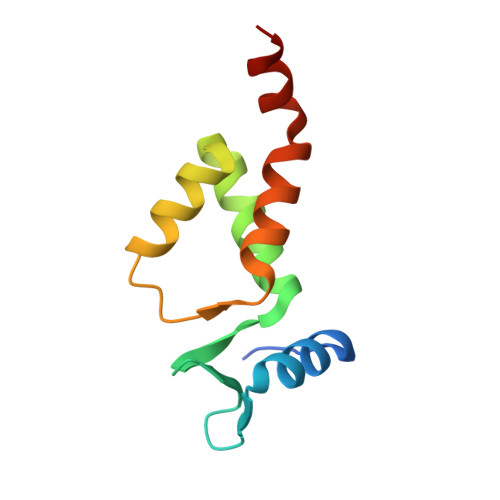
    The retroviral integrase (IN) carries out the integration of a dsDNA copy of the viral genome into the host DNA, an essential step for viral replication. All IN proteins have three general domains, the N-terminal domain (NTD), the catalytic core domain, and the C-terminal domain. The NTD includes an HHCC zinc finger-like motif, which is conserved in all retroviral IN proteins. Two crystal structures of Moloney murine leukemia virus (M-MuLV) IN N-terminal region (NTR) constructs that both include an N-terminal extension domain (NED, residues 1-44) and an HHCC zinc-finger NTD (residues 45-105), in two crystal forms are reported. The structures of IN NTR constructs encoding residues 1-105 (NTR 1-105 ) and 8-105 (NTR 8-105 ) were determined at 2.7 and 2.15 Å resolution, respectively and belong to different space groups. While both crystal forms have similar protomer structures, NTR 1-105 packs as a dimer and NTR 8-105 packs as a tetramer in the asymmetric unit. The structure of the NED consists of three anti-parallel β-strands and an α-helix, similar to the NED of prototype foamy virus (PFV) IN. These three β-strands form an extended β-sheet with another β-strand in the HHCC Zn 2+ binding domain, which is a unique structural feature for the M-MuLV IN. The HHCC Zn 2+ binding domain structure is similar to that in HIV and PFV INs, with variations within the loop regions. Differences between the PFV and MLV IN NEDs localize at regions identified to interact with the PFV LTR and are compared with established biochemical and virological data for M-MuLV. Proteins 2017; 85:647-656. © 2016 Wiley Periodicals, Inc.


  • Organizational Affiliation
    • Center for Advanced Biotechnology and Medicine, Rutgers, The State University of New Jersey, Piscataway, New Jersey, 08854.

Macromolecule Content 

  • Total Structure Weight: 48.83 kDa 
  • Atom Count: 3,243 
  • Modeled Residue Count: 372 
  • Deposited Residue Count: 400 
  • Unique protein chains: 1

Macromolecules

Find similar proteins by:|  3D Structure
Entity ID: 1
MoleculeChains  Sequence LengthOrganismDetailsImage
Integrase p46
A, B, C, D
100Moloney murine leukemia virus isolate ShinnickMutation(s): 0 
Gene Names: gag-pol
UniProt
Find proteins for P03355 (Moloney murine leukemia virus (isolate Shinnick))
Explore P03355 
Go to UniProtKB:  P03355
Entity Groups
Sequence Clusters30% Identity50% Identity70% Identity90% Identity95% Identity100% Identity
UniProt GroupP03355
Sequence Annotations
Expand
Reference Sequence

Small Molecules

Ligands 3 Unique
IDChains Name / Formula / InChI Key2D Diagram3D Interactions
DTT

Query on DTT


Download:Ideal Coordinates CCD File
G [auth A]
H [auth A]
I [auth A]
L [auth B]
M [auth B]
G [auth A],
H [auth A],
I [auth A],
L [auth B],
M [auth B],
N [auth B],
P [auth C],
Q [auth C],
S [auth D]
2,3-DIHYDROXY-1,4-DITHIOBUTANE
C4 H10 O2 S2
VHJLVAABSRFDPM-IMJSIDKUSA-N
ZN

Query on ZN


Download:Ideal Coordinates CCD File
E [auth A],
J [auth B],
O [auth C],
T [auth D]
ZINC ION
Zn
PTFCDOFLOPIGGS-UHFFFAOYSA-N
ACT

Query on ACT


Download:Ideal Coordinates CCD File
F [auth A],
K [auth B],
R [auth D]
ACETATE ION
C2 H3 O2
QTBSBXVTEAMEQO-UHFFFAOYSA-M

Experimental Data & Validation

Experimental Data

  • Method: X-RAY DIFFRACTION
  • Resolution: 2.15 Å
  • R-Value Free:  0.265 (Depositor), 0.268 (DCC) 
  • R-Value Work:  0.234 (Depositor), 0.238 (DCC) 
  • R-Value Observed: 0.236 (Depositor) 
Space Group: P 1 21 1
Unit Cell:
Length ( Å )Angle ( ˚ )
a = 44.659α = 90
b = 38.44β = 91.7
c = 135.275γ = 90
Software Package:
Software NamePurpose
PHENIXrefinement
PDB_EXTRACTdata extraction
CBASSdata collection
HKL-2000data reduction
HKL-2000data scaling
PHASERphasing

Structure Validation

View Full Validation Report


Entry History 

Revision History  (Full details and data files)

  • Version 1.0: 2014-02-05
    Type: Initial release
  • Version 1.1: 2017-02-22
    Changes: Database references
  • Version 1.2: 2022-08-24
    Changes: Database references, Derived calculations
  • Version 1.3: 2023-09-20
    Changes: Data collection, Refinement description