4NZC | pdb_00004nzc

Crystal structure of Chitinase D from Serratia proteamaculans at 1.45 Angstrom resolution


Experimental Data Snapshot

  • Method: X-RAY DIFFRACTION
  • Resolution: 1.45 Å
  • R-Value Free: 
    0.213 (Depositor), 0.219 (DCC) 
  • R-Value Work: 
    0.199 (Depositor), 0.207 (DCC) 
  • R-Value Observed: 
    0.200 (Depositor) 

Starting Model: experimental
View more details

wwPDB Validation 3D Report Full Report

Validation slider image for 4NZC

This is version 1.3 of the entry. See complete history

Literature

Structure of chitinase D from Serratia proteamaculans reveals the structural basis of its dual action of hydrolysis and transglycosylation

Madhuprakash, J.Singh, A.Kumar, S.Sinha, M.Kaur, P.Sharma, S.Podile, A.R.Singh, T.P.

(2013) Int J Biochem Mol Biol 4: 166-178

  • Primary Citation Related Structures: 
    4NZC

  • PubMed Abstract: 

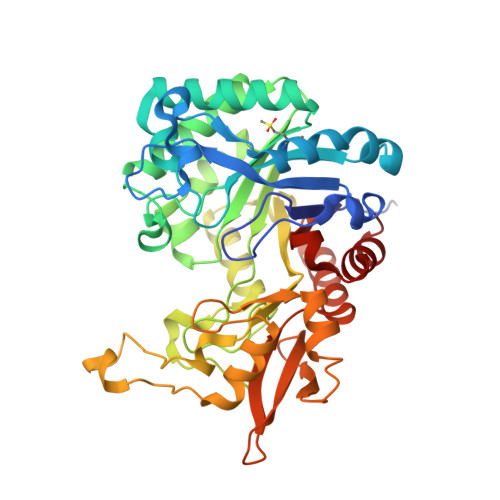
    Chitinases are known to hydrolyze chitin polymers into smaller chitooligosaccharides. Chitinase from bacterium Serratia proteamaculans (SpChiD) is found to exhibit both hydrolysis and transglycosylation activities. SpChiD belongs to family 18 of glycosyl hydrolases (GH-18). The recombinant SpChiD was crystallized and its three-dimensional structure was determined at 1.49 Å resolution. The structure was refined to an R-factor of 16.2%. SpChiD consists of 406 amino acid residues. The polypeptide chain of SpChiD adopts a (β/α)8 triosephosphate isomerase (TIM) barrel structure. SpChiD contains three acidic residues, Asp149, Asp151 and Glu153 as part of its catalytic scheme. While both Asp149 and Glu153 adopt single conformations, Asp151 is observed in two conformations. The substrate binding cleft is partially obstructed by a protruding loop, Asn30 - Asp42 causing a considerable reduction in the number of available subsites in the substrate binding site. The positioning of loop, Asn30 - Asp42 appears to be responsible for the transglycosylation activity. The structure determination indicated the presence of sulfone Met89 (SMet89). The sulfone methionine residue is located on the surface of the protein at a site where extra domain is attached in other chitinases. This is the first structure of a single domain chitinase with hydrolytic and transglycosylation activities.


  • Organizational Affiliation
    • Department of Plant Sciences, School of Life Sciences, University of Hyderabad Hyderabad, India.

Macromolecule Content 

  • Total Structure Weight: 44.36 kDa 
  • Atom Count: 3,761 
  • Modeled Residue Count: 398 
  • Deposited Residue Count: 398 
  • Unique protein chains: 1

Macromolecules

Find similar proteins by:|  3D Structure
Entity ID: 1
MoleculeChains  Sequence LengthOrganismDetailsImage
Glycoside hydrolase family 18398Serratia proteamaculans 568Mutation(s): 0 
Gene Names: Spro_2725
EC: 3.2.1.14
UniProt
Find proteins for A8GFD6 (Serratia proteamaculans (strain 568))
Explore A8GFD6 
Go to UniProtKB:  A8GFD6
Entity Groups
Sequence Clusters30% Identity50% Identity70% Identity90% Identity95% Identity100% Identity
UniProt GroupA8GFD6
Sequence Annotations
Expand
Reference Sequence

Experimental Data & Validation

Experimental Data

  • Method: X-RAY DIFFRACTION
  • Resolution: 1.45 Å
  • R-Value Free:  0.213 (Depositor), 0.219 (DCC) 
  • R-Value Work:  0.199 (Depositor), 0.207 (DCC) 
  • R-Value Observed: 0.200 (Depositor) 
Space Group: P 2 21 21
Unit Cell:
Length ( Å )Angle ( ˚ )
a = 59.334α = 90
b = 75.276β = 90
c = 86.713γ = 90
Software Package:
Software NamePurpose
HKL-2000data collection
AMoREphasing
REFMACrefinement
HKL-2000data reduction
SCALEPACKdata scaling

Structure Validation

View Full Validation Report


Entry History 

Deposition Data

Revision History  (Full details and data files)

  • Version 1.0: 2014-01-01
    Type: Initial release
  • Version 1.1: 2014-11-19
    Changes: Database references
  • Version 1.2: 2023-11-08
    Changes: Data collection, Database references, Derived calculations, Refinement description
  • Version 1.3: 2024-10-30
    Changes: Structure summary