4NYO | pdb_00004nyo

The 1.8 Angstrom Crystal Structure of the Periplasmic Divalent Cation Tolerance Protein Cuta from Pyrococcus Horikoshii OT3


Experimental Data Snapshot

  • Method: X-RAY DIFFRACTION
  • Resolution: 1.80 Å
  • R-Value Free: 
    0.225 (Depositor), 0.225 (DCC) 
  • R-Value Work: 
    0.189 (Depositor), 0.189 (DCC) 
  • R-Value Observed: 
    0.189 (Depositor) 

Starting Model: experimental
View more details

wwPDB Validation 3D Report Full Report

Validation slider image for 4NYO

This is version 1.4 of the entry. See complete history

Literature

The structures of the CutA1 proteins from Thermus thermophilus and Pyrococcus horikoshii: characterization of metal-binding sites and metal-induced assembly

Bagautdinov, B.

(2014) Acta Crystallogr F Struct Biol Commun 70: 404-413

  • DOI: https://doi.org/10.1107/S2053230X14003422
  • Primary Citation Related Structures: 
    1NZA, 1V6H, 4NYO

  • PubMed Abstract: 

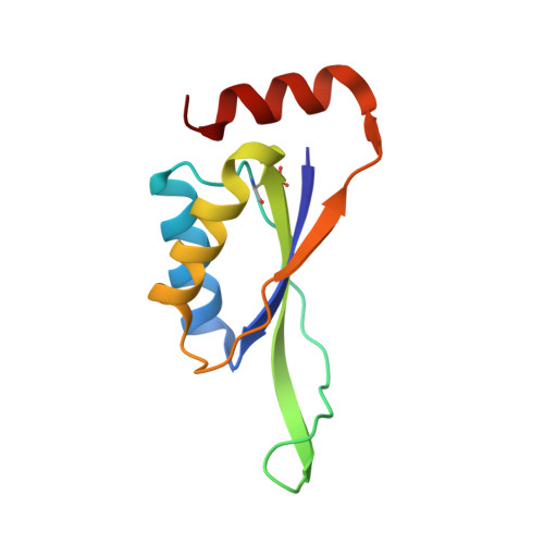
    CutA1 (copper tolerance A1) is a widespread cytoplasmic protein found in archaea, bacteria, plants and animals, including humans. In Escherichia coli it is implicated in divalent metal tolerance, while the mammalian CutA1 homologue has been proposed to mediate brain enzyme acetylcholinesterase activity and copper homeostasis. The X-ray structures of CutA1 from the thermophilic bacterium Thermus thermophilus (TtCutA1) with and without bound Na(+) at 1.7 and 1.9 Å resolution, respectively, and from the hyperthermophilic archaeon Pyrococcus horikoshii (PhCutA1) in complex with Na(+) at 1.8 Å resolution have been determined. Both are short and rigid proteins of about 12 kDa that form intertwined compact trimers in the crystal and solution. The main difference in the structures is a wide-type β-bulge on top of the TtCutA1 trimer. It affords a mechanism for lodging a single-residue insertion in the middle of β2 while preserving the interprotomer main-chain hydrogen-bonding network. The liganded forms of the proteins provide new structural information about the metal-binding sites and CutA1 assembly. The Na(+)-TtCutA1 structure unveils a dodecameric assembly with metal ions in the trimer-trimer interfaces and the lateral clefts of the trimer. For Na(+)-PhCutA1, the metal ion associated with six waters in an octahedral geometry. The structures suggest that CutA1 may contribute to regulating intracellular metal homeostasis through various binding modes.


  • Organizational Affiliation
    • Japan Synchrotron Radiation Research Institute (JASRI/SPring-8), 1-1-1 Kouto, Sayo, Hyogo 679-5198, Japan.

Macromolecule Content 

  • Total Structure Weight: 75.7 kDa 
  • Atom Count: 6,012 
  • Modeled Residue Count: 612 
  • Deposited Residue Count: 612 
  • Unique protein chains: 1

Macromolecules

Find similar proteins by:|  3D Structure
Entity ID: 1
MoleculeChains  Sequence LengthOrganismDetailsImage
Divalent-cation tolerance protein CutA
A, B, C, D, E
A, B, C, D, E, F
102Pyrococcus horikoshii OT3Mutation(s): 0 
Gene Names: cutAPH0992
UniProt
Find proteins for O58720 (Pyrococcus horikoshii (strain ATCC 700860 / DSM 12428 / JCM 9974 / NBRC 100139 / OT-3))
Explore O58720 
Go to UniProtKB:  O58720
Entity Groups
Sequence Clusters30% Identity50% Identity70% Identity90% Identity95% Identity100% Identity
UniProt GroupO58720
Sequence Annotations
Expand
Reference Sequence

Small Molecules

Ligands 4 Unique
IDChains Name / Formula / InChI Key2D Diagram3D Interactions
MES

Query on MES


Download:Ideal Coordinates CCD File
I [auth A],
L [auth B],
T [auth D],
V [auth F]
2-(N-MORPHOLINO)-ETHANESULFONIC ACID
C6 H13 N O4 S
SXGZJKUKBWWHRA-UHFFFAOYSA-N
SO4

Query on SO4


Download:Ideal Coordinates CCD File
N [auth C],
S [auth D]
SULFATE ION
O4 S
QAOWNCQODCNURD-UHFFFAOYSA-L
CL

Query on CL


Download:Ideal Coordinates CCD File
H [auth A]
J [auth B]
K [auth B]
M [auth C]
P [auth D]
H [auth A],
J [auth B],
K [auth B],
M [auth C],
P [auth D],
Q [auth D],
R [auth D],
U [auth E]
CHLORIDE ION
Cl
VEXZGXHMUGYJMC-UHFFFAOYSA-M
NA

Query on NA


Download:Ideal Coordinates CCD File
G [auth A],
O [auth D]
SODIUM ION
Na
FKNQFGJONOIPTF-UHFFFAOYSA-N
Modified Residues  1 Unique
IDChains TypeFormula2D DiagramParent
CSD
Query on CSD
A, B, C, D, E
A, B, C, D, E, F
L-PEPTIDE LINKINGC3 H7 N O4 SCYS

Experimental Data & Validation

Experimental Data

  • Method: X-RAY DIFFRACTION
  • Resolution: 1.80 Å
  • R-Value Free:  0.225 (Depositor), 0.225 (DCC) 
  • R-Value Work:  0.189 (Depositor), 0.189 (DCC) 
  • R-Value Observed: 0.189 (Depositor) 
Space Group: P 21 21 21
Unit Cell:
Length ( Å )Angle ( ˚ )
a = 93.085α = 90
b = 124.25β = 90
c = 50.458γ = 90
Software Package:
Software NamePurpose
CrystalCleardata collection
MOLREPphasing
CNSrefinement
HKL-2000data reduction
SCALEPACKdata scaling

Structure Validation

View Full Validation Report


Entry History 

Deposition Data

Revision History  (Full details and data files)

  • Version 1.0: 2014-01-01
    Type: Initial release
  • Version 1.1: 2014-04-16
    Changes: Database references
  • Version 1.2: 2014-11-12
    Changes: Other
  • Version 1.3: 2023-11-08
    Changes: Data collection, Database references, Derived calculations, Refinement description
  • Version 1.4: 2024-11-06
    Changes: Structure summary