


Bond length analysis of the PqqC Y175F mutant structure shows evidence for bound PQQ in the reduced form
Fisher, S.J., Puehringer, S.To be published.
Experimental Data Snapshot
Starting Model: experimental
View more details
Entity ID: 1 | |||||
|---|---|---|---|---|---|
| Molecule | Chains | Sequence Length | Organism | Details | Image |
| Pyrroloquinoline-quinone synthase | 258 | Klebsiella pneumoniae subsp. pneumoniae MGH 78578 | Mutation(s): 1 Gene Names: pqqC, KPN78578_17810, KPN_01811 EC: 1.3.3.11 | 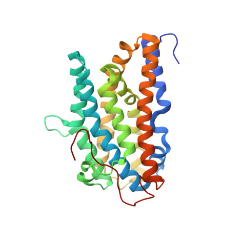 | |
UniProt | |||||
Entity Groups | |||||
| Sequence Clusters | 30% Identity50% Identity70% Identity90% Identity95% Identity100% Identity | ||||
| UniProt Group | A6T9H1 | ||||
Sequence AnnotationsExpand | |||||
Reference Sequence | |||||
| Ligands 3 Unique | |||||
|---|---|---|---|---|---|
| ID | Chains | Name / Formula / InChI Key | 2D Diagram | 3D Interactions | |
| PQQ Download:Ideal Coordinates CCD File | C [auth A], J [auth B] | PYRROLOQUINOLINE QUINONE C14 H6 N2 O8 MMXZSJMASHPLLR-UHFFFAOYSA-N |  | ||
| GOL Download:Ideal Coordinates CCD File | D [auth A] E [auth A] G [auth A] H [auth A] I [auth A] | GLYCEROL C3 H8 O3 PEDCQBHIVMGVHV-UHFFFAOYSA-N |  | ||
| CL Download:Ideal Coordinates CCD File | F [auth A], M [auth B] | CHLORIDE ION Cl VEXZGXHMUGYJMC-UHFFFAOYSA-M |  | ||
| Length ( Å ) | Angle ( ˚ ) |
|---|---|
| a = 70.998 | α = 90 |
| b = 116.02 | β = 90 |
| c = 67.52 | γ = 90 |
| Software Name | Purpose |
|---|---|
| MX | data collection |
| PHENIX | model building |
| SHELXL-97 | refinement |
| XDS | data reduction |
| SCALA | data scaling |
| PHENIX | phasing |


