4NX2 | pdb_00004nx2

Crystal structure of DCYRS complexed with DCY


Experimental Data Snapshot

  • Method: X-RAY DIFFRACTION
  • Resolution: 2.00 Å
  • R-Value Free: 
    0.220 (Depositor), 0.231 (DCC) 
  • R-Value Work: 
    0.172 (Depositor), 0.180 (DCC) 
  • R-Value Observed: 
    0.174 (Depositor) 

wwPDB Validation 3D Report Full Report

Validation slider image for 4NX2

Ligand Structure Quality Assessment 


This is version 1.2 of the entry. See complete history

Literature

Significant expansion of fluorescent protein sensing ability through the genetic incorporation of superior photo-induced electron-transfer quenchers.

Liu, X.Jiang, L.Li, J.Wang, L.Yu, Y.Zhou, Q.Lv, X.Gong, W.Lu, Y.Wang, J.

(2014) J Am Chem Soc 136: 13094-13097

  • DOI: https://doi.org/10.1021/ja505219r
  • Primary Citation Related Structures: 
    4NX2, 4NXB, 4NXE, 4NXF, 4NXG

  • PubMed Abstract: 

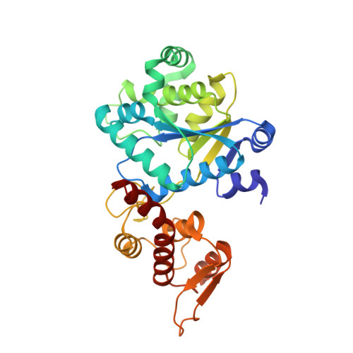
    Photo-induced electron transfer (PET) is ubiquitous for photosynthesis and fluorescent sensor design. However, genetically coded PET sensors are underdeveloped, due to the lack of methods to site-specifically install PET probes on proteins. Here we describe a family of acid and Mn(III) turn-on fluorescent protein (FP) sensors, named iLovU, based on PET and the genetic incorporation of superior PET quenchers in the fluorescent flavoprotein iLov. Using the iLovU PET sensors, we monitored the cytoplasmic acidification process, and achieved Mn(III) fluorescence sensing for the first time. The iLovU sensors should be applicable for studying pH changes in living cells, monitoring biogentic Mn(III) in the environment, and screening for efficient manganese peroxidase, which is highly desirable for lignin degradation and biomass conversion. Our work establishes a platform for many more protein PET sensors, facilitates the de novo design of metalloenzymes harboring redox active residues, and expands our ability to probe protein conformational dynamics.


  • Organizational Affiliation
    • Laboratory of RNA Biology, Institute of Biophysics, Chinese Academy of Sciences , Chaoyang District, Beijing, 100101, China.

Macromolecule Content 

  • Total Structure Weight: 35.96 kDa 
  • Atom Count: 2,721 
  • Modeled Residue Count: 313 
  • Deposited Residue Count: 313 
  • Unique protein chains: 1

Macromolecules

Find similar proteins by:|  3D Structure
Entity ID: 1
MoleculeChains  Sequence LengthOrganismDetailsImage
Tyrosine--tRNA ligase313Methanocaldococcus jannaschii DSM 2661Mutation(s): 8 
Gene Names: tyrSMJ0389
EC: 6.1.1.1
UniProt
Find proteins for Q57834 (Methanocaldococcus jannaschii (strain ATCC 43067 / DSM 2661 / JAL-1 / JCM 10045 / NBRC 100440))
Explore Q57834 
Go to UniProtKB:  Q57834
Entity Groups
Sequence Clusters30% Identity50% Identity70% Identity90% Identity95% Identity100% Identity
UniProt GroupQ57834
Sequence Annotations
Expand
Reference Sequence

Small Molecules

Ligands 1 Unique
IDChains Name / Formula / InChI Key2D Diagram3D Interactions
2LT

Query on 2LT


Download:Ideal Coordinates CCD File
B [auth A]3,5-dichloro-L-tyrosine
C9 H9 Cl2 N O3
MPHURJQUHZHALJ-ZETCQYMHSA-N

Experimental Data & Validation

Experimental Data

  • Method: X-RAY DIFFRACTION
  • Resolution: 2.00 Å
  • R-Value Free:  0.220 (Depositor), 0.231 (DCC) 
  • R-Value Work:  0.172 (Depositor), 0.180 (DCC) 
  • R-Value Observed: 0.174 (Depositor) 
Space Group: P 21 21 2
Unit Cell:
Length ( Å )Angle ( ˚ )
a = 53.099α = 90
b = 160.955β = 90
c = 39.009γ = 90
Software Package:
Software NamePurpose
HKL-2000data collection
MOLREPphasing
PHENIXrefinement
HKL-2000data reduction
HKL-2000data scaling

Structure Validation

View Full Validation Report


Ligand Structure Quality Assessment 


Entry History 

Deposition Data

Revision History  (Full details and data files)

  • Version 1.0: 2014-09-24
    Type: Initial release
  • Version 1.1: 2022-08-24
    Changes: Database references, Derived calculations
  • Version 1.2: 2024-05-29
    Changes: Data collection