4NV6

C212A mutant of Synechococcus VKOR


Experimental Data Snapshot

  • Method: X-RAY DIFFRACTION
  • Resolution: 4.19 Å
  • R-Value Free: 0.331 
  • R-Value Work: 0.299 
  • R-Value Observed: 0.302 

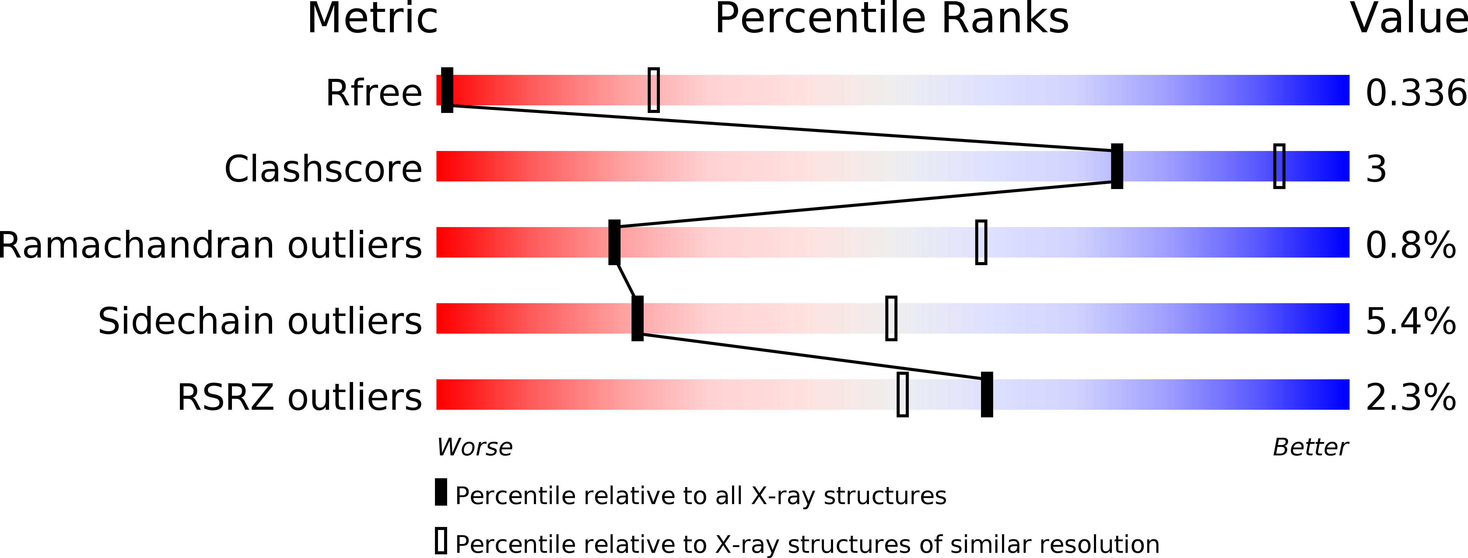
wwPDB Validation   3D Report Full Report


Ligand Structure Quality Assessment 


This is version 1.1 of the entry. See complete history


Literature

Structures of an intramembrane vitamin K epoxide reductase homolog reveal control mechanisms for electron transfer.

Liu, S.Cheng, W.Fowle Grider, R.Shen, G.Li, W.

(2014) Nat Commun 5: 3110-3110

  • DOI: https://doi.org/10.1038/ncomms4110
  • Primary Citation of Related Structures:  
    4NV2, 4NV5, 4NV6

  • PubMed Abstract: 

    The intramembrane vitamin K epoxide reductase (VKOR) supports blood coagulation in humans and is the target of the anticoagulant warfarin. VKOR and its homologues generate disulphide bonds in organisms ranging from bacteria to humans. Here, to better understand the mechanism of VKOR catalysis, we report two crystal structures of a bacterial VKOR captured in different reaction states. These structures reveal a short helix at the hydrophobic active site of VKOR that alters between wound and unwound conformations. Motions of this 'horizontal helix' promote electron transfer by regulating the positions of two cysteines in an adjacent loop. Winding of the helix separates these 'loop cysteines' to prevent backward electron flow. Despite these motions, hydrophobicity at the active site is maintained to facilitate VKOR catalysis. Biochemical experiments suggest that several warfarin-resistant mutations act by changing the conformation of the horizontal helix. Taken together, these studies provide a comprehensive understanding of VKOR function.


  • Organizational Affiliation

    1] Department of Biochemistry and Molecular Biophysics, Washington University School of Medicine, St Louis, Missouri 63110, USA [2].


Macromolecules
Find similar proteins by:  (by identity cutoff)  |  3D Structure
Entity ID: 1
MoleculeChains Sequence LengthOrganismDetailsImage
VKORC1/thioredoxin domain protein291Synechococcus sp. JA-2-3B'a(2-13)Mutation(s): 1 
Gene Names: CYB_2278
EC: 1.17.4
Membrane Entity: Yes 
UniProt
Find proteins for Q2JJF6 (Synechococcus sp. (strain JA-2-3B'a(2-13)))
Explore Q2JJF6 
Go to UniProtKB:  Q2JJF6
Entity Groups  
Sequence Clusters30% Identity50% Identity70% Identity90% Identity95% Identity100% Identity
UniProt GroupQ2JJF6
Sequence Annotations
Expand
  • Reference Sequence
Small Molecules
Ligands 1 Unique
IDChains Name / Formula / InChI Key2D Diagram3D Interactions
U10
Query on U10

Download Ideal Coordinates CCD File 
B [auth A]UBIQUINONE-10
C59 H90 O4
ACTIUHUUMQJHFO-UPTCCGCDSA-N
Experimental Data & Validation

Experimental Data

  • Method: X-RAY DIFFRACTION
  • Resolution: 4.19 Å
  • R-Value Free: 0.331 
  • R-Value Work: 0.299 
  • R-Value Observed: 0.302 
  • Space Group: P 61
Unit Cell:
Length ( Å )Angle ( ˚ )
a = 139.563α = 90
b = 139.563β = 90
c = 68.724γ = 120
Software Package:
Software NamePurpose
HKL-2000data collection
PARROTphasing
PHENIXrefinement
HKL-2000data reduction
HKL-2000data scaling

Structure Validation

View Full Validation Report


Ligand Structure Quality Assessment 


Entry History 

Deposition Data

Revision History  (Full details and data files)

  • Version 1.0: 2014-02-12
    Type: Initial release
  • Version 1.1: 2024-10-16
    Changes: Data collection, Database references, Derived calculations, Structure summary