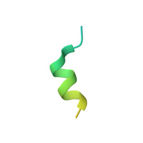
4NUU

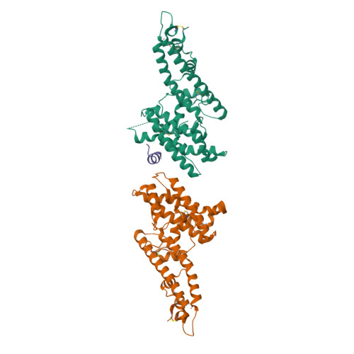
Heterotrimer structure of Region II from Plasmodium vivax Duffy Binding Protein (PvDBP) bound to the ectodomain of the Duffy Antigen Receptor for Chemokines (DARC)


Experimental Data Snapshot

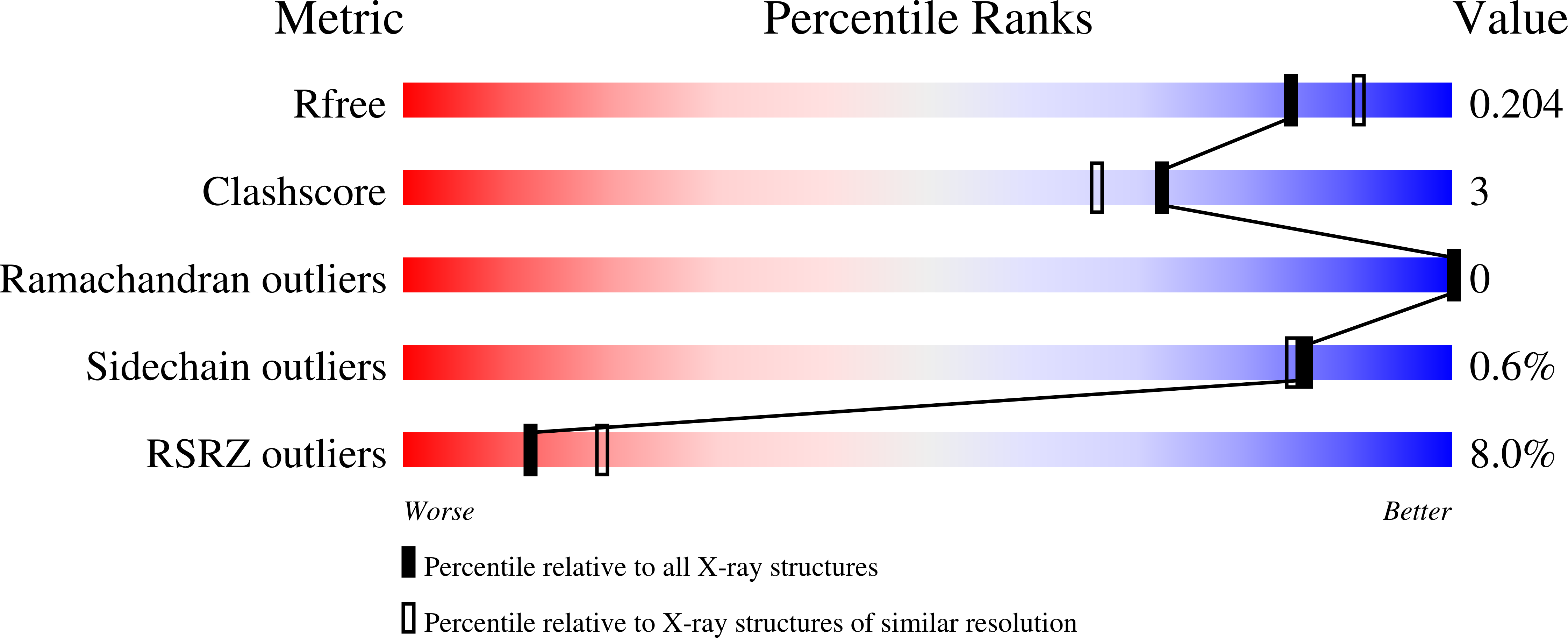
  • Method: X-RAY DIFFRACTION
  • Resolution: 1.95 Å
  • R-Value Free: 0.202 
  • R-Value Work: 0.165 
  • R-Value Observed: 0.167 

wwPDB Validation   3D Report Full Report


This is version 1.3 of the entry. See complete history


Literature

Red Blood Cell Invasion by Plasmodium vivax: Structural Basis for DBP Engagement of DARC.

Batchelor, J.D.Malpede, B.M.Omattage, N.S.Dekoster, G.T.Henzler-Wildman, K.A.Tolia, N.H.

(2014) PLoS Pathog 10: e1003869-e1003869

  • DOI: https://doi.org/10.1371/journal.ppat.1003869
  • Primary Citation of Related Structures:  
    4NUU, 4NUV

  • PubMed Abstract: 

    Plasmodium parasites use specialized ligands which bind to red blood cell (RBC) receptors during invasion. Defining the mechanism of receptor recognition is essential for the design of interventions against malaria. Here, we present the structural basis for Duffy antigen (DARC) engagement by P. vivax Duffy binding protein (DBP). We used NMR to map the core region of the DARC ectodomain contacted by the receptor binding domain of DBP (DBP-RII) and solved two distinct crystal structures of DBP-RII bound to this core region of DARC. Isothermal titration calorimetry studies show these structures are part of a multi-step binding pathway, and individual point mutations of residues contacting DARC result in a complete loss of RBC binding by DBP-RII. Two DBP-RII molecules sandwich either one or two DARC ectodomains, creating distinct heterotrimeric and heterotetrameric architectures. The DARC N-terminus forms an amphipathic helix upon DBP-RII binding. The studies reveal a receptor binding pocket in DBP and critical contacts in DARC, reveal novel targets for intervention, and suggest that targeting the critical DARC binding sites will lead to potent disruption of RBC engagement as complex assembly is dependent on DARC binding. These results allow for models to examine inter-species infection barriers, Plasmodium immune evasion mechanisms, P. knowlesi receptor-ligand specificity, and mechanisms of naturally acquired P. vivax immunity. The step-wise binding model identifies a possible mechanism by which signaling pathways could be activated during invasion. It is anticipated that the structural basis of DBP host-cell engagement will enable development of rational therapeutics targeting this interaction.


  • Organizational Affiliation

    Department of Molecular Microbiology and Microbial Pathogenesis, Washington University School of Medicine, Saint Louis, Missouri, United States of America.


Macromolecules
Find similar proteins by:  (by identity cutoff)  |  3D Structure
Entity ID: 1
MoleculeChains Sequence LengthOrganismDetailsImage
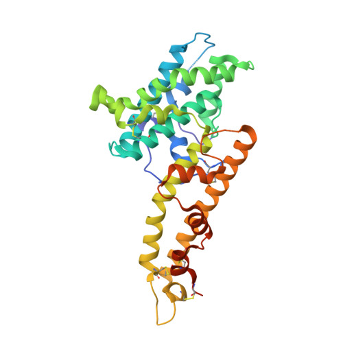
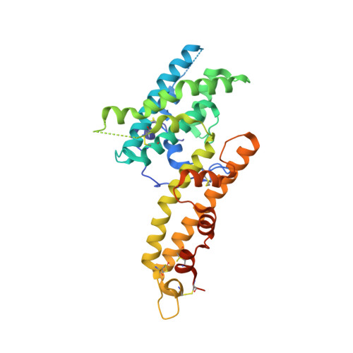
Duffy receptor
A, B
317Plasmodium vivax Sal-1Mutation(s): 0 
Gene Names: PVDR
UniProt
Find proteins for P22290 (Plasmodium vivax (strain Salvador I))
Explore P22290 
Go to UniProtKB:  P22290
Entity Groups  
Sequence Clusters30% Identity50% Identity70% Identity90% Identity95% Identity100% Identity
UniProt GroupP22290
Sequence Annotations
Expand
  • Reference Sequence
Find similar proteins by:  (by identity cutoff)  |  3D Structure
Entity ID: 2
MoleculeChains Sequence LengthOrganismDetailsImage
Duffy antigen/chemokine receptor32Homo sapiensMutation(s): 0 
Gene Names: DARCACKR1FYGPD
UniProt & NIH Common Fund Data Resources
Find proteins for Q16570 (Homo sapiens)
Explore Q16570 
Go to UniProtKB:  Q16570
PHAROS:  Q16570
GTEx:  ENSG00000213088 
Entity Groups  
Sequence Clusters30% Identity50% Identity70% Identity90% Identity95% Identity100% Identity
UniProt GroupQ16570
Sequence Annotations
Expand
  • Reference Sequence
Experimental Data & Validation

Experimental Data

  • Method: X-RAY DIFFRACTION
  • Resolution: 1.95 Å
  • R-Value Free: 0.202 
  • R-Value Work: 0.165 
  • R-Value Observed: 0.167 
  • Space Group: P 1 21 1
Unit Cell:
Length ( Å )Angle ( ˚ )
a = 59.59α = 90
b = 66.99β = 102.11
c = 97.92γ = 90
Software Package:
Software NamePurpose
Blu-Icedata collection
PHASERphasing
PHENIXrefinement
XDSdata reduction
XDSdata scaling

Structure Validation

View Full Validation Report


Entry History 

Deposition Data

  • Released Date: 2014-02-05 
  • Deposition Author(s): Tolia, N.H.

Revision History  (Full details and data files)

  • Version 1.0: 2014-02-05
    Type: Initial release
  • Version 1.1: 2017-11-22
    Changes: Refinement description
  • Version 1.2: 2024-03-13
    Changes: Data collection, Database references, Source and taxonomy
  • Version 1.3: 2024-10-30
    Changes: Structure summary