4NSV | pdb_00004nsv

Lysobacter enzymogenes lysc endoproteinase K30R mutant covalently inhibited by TLCK


Experimental Data Snapshot

  • Method: X-RAY DIFFRACTION
  • Resolution: 0.90 Å
  • R-Value Free: 
    0.158 (Depositor), 0.142 (DCC) 
  • R-Value Work: 
    0.138 (Depositor) 
  • R-Value Observed: 
    0.139 (Depositor) 

Starting Model: experimental
View more details

wwPDB Validation 3D Report Full Report

Validation slider image for 4NSV

Ligand Structure Quality Assessment 


This is version 1.5 of the entry. See complete history

Literature

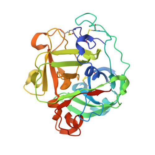
Atomic resolution structure of a lysine-specific endoproteinase from Lysobacter enzymogenes suggests a hydroxyl group bound to the oxyanion hole.

Asztalos, P.Muller, A.Holke, W.Sobek, H.Rudolph, M.G.

(2014) Acta Crystallogr D Biol Crystallogr 70: 1832-1843

  • DOI: https://doi.org/10.1107/S1399004714008463
  • Primary Citation Related Structures: 
    4NSV, 4NSY

  • PubMed Abstract: 

    Lysobacter enzymogenes lysyl endoproteinase (LysC) is a trypsin-type serine protease with a high pH optimum that hydrolyses all Lys-Xaa peptide bonds. The high specificity of LysC renders it useful for biotechnological purposes. The K30R variant of a related lysyl endoproteinase from Achromobacter lyticus has favourable enzymatic properties that might be transferrable to LysC. To visualize structural differences in the substrate-binding sites, the crystal structures of wild-type and the K30R variant of LysC were determined. The mutation is located at a distance of 12 Å from the catalytic triad and subtly changes the surface properties of the substrate-binding site. The high pH optimum of LysC can be attributed to electrostatic effects of an aromatic Tyr/His stack on the catalytic aspartate and is a general feature of this enzyme subfamily. LysC crystals in complex with the covalent inhibitor N(α)-p-tosyl-lysyl chloromethylketone yielded data to 1.1 and 0.9 Å resolution, resulting in unprecedented precision of the active and substrate-binding sites for this enzyme subfamily. Error estimates on bond lengths and difference electron density indicate that instead of the expected oxyanion a hydroxyl group binds to the partially solvent-exposed oxyanion hole. Protonation of the alkoxide catalytic intermediate might be a recurring feature during serine protease catalysis.


  • Organizational Affiliation
    • Roche Diagnostics GmbH, Nonnenwald 2, 82377 Penzberg, Germany.

Macromolecule Content 

  • Total Structure Weight: 58.49 kDa 
  • Atom Count: 5,075 
  • Modeled Residue Count: 528 
  • Deposited Residue Count: 550 
  • Unique protein chains: 1

Macromolecules

Find similar proteins by:|  3D Structure
Entity ID: 1
MoleculeChains  Sequence LengthOrganismDetailsImage
Lysyl endopeptidase
A, B
275Lysobacter enzymogenesMutation(s): 1 
EC: 3.4.21.50
UniProt
Find proteins for Q7M135 (Lysobacter enzymogenes)
Explore Q7M135 
Go to UniProtKB:  Q7M135
Entity Groups
Sequence Clusters30% Identity50% Identity70% Identity90% Identity95% Identity100% Identity
UniProt GroupQ7M135
Sequence Annotations
Expand
Reference Sequence

Small Molecules

Experimental Data & Validation

Experimental Data

  • Method: X-RAY DIFFRACTION
  • Resolution: 0.90 Å
  • R-Value Free:  0.158 (Depositor), 0.142 (DCC) 
  • R-Value Work:  0.138 (Depositor) 
  • R-Value Observed: 0.139 (Depositor) 
Space Group: P 1 21 1
Unit Cell:
Length ( Å )Angle ( ˚ )
a = 39.605α = 90
b = 135.582β = 98.01
c = 45.553γ = 90
Software Package:
Software NamePurpose
PHASERphasing
PHENIXrefinement
XDSdata reduction
XSCALEdata scaling

Structure Validation

View Full Validation Report


Ligand Structure Quality Assessment 


Entry History 

Deposition Data

Revision History  (Full details and data files)

  • Version 1.0: 2014-04-23
    Type: Initial release
  • Version 1.1: 2014-07-09
    Changes: Database references
  • Version 1.2: 2014-09-24
    Changes: Database references
  • Version 1.3: 2017-11-22
    Changes: Refinement description
  • Version 1.4: 2023-09-20
    Changes: Data collection, Database references, Derived calculations, Refinement description
  • Version 1.5: 2024-11-20
    Changes: Structure summary