4NSJ | pdb_00004nsj

Carboplatin binding to HEWL in 2M NH4formate, 0.1M HEPES at pH 7.5


Experimental Data Snapshot

  • Method: X-RAY DIFFRACTION
  • Resolution: 1.70 Å
  • R-Value Free: 
    0.258 (Depositor), 0.265 (DCC) 
  • R-Value Work: 
    0.199 (Depositor), 0.206 (DCC) 
  • R-Value Observed: 
    0.202 (Depositor) 

Starting Model: experimental
View more details

wwPDB Validation 3D Report Full Report

Validation slider image for 4NSJ

This is version 1.3 of the entry. See complete history

Literature

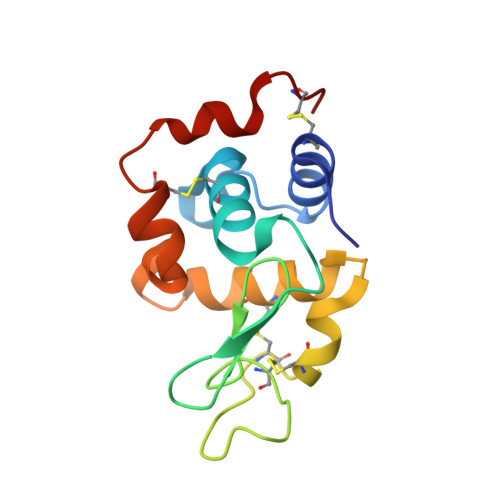
Carboplatin binding to histidine.

Tanley, S.W.Diederichs, K.Kroon-Batenburg, L.M.Levy, C.Schreurs, A.M.Helliwell, J.R.

(2014) Acta Crystallogr F Struct Biol Commun 70: 1135-1142

  • DOI: https://doi.org/10.1107/S2053230X14016161
  • Primary Citation Related Structures: 
    4LT0, 4LT3, 4NSG, 4NSH, 4NSI, 4NSJ

  • PubMed Abstract: 

    Carboplatin is a second-generation platinum anticancer agent used for the treatment of a variety of cancers. Previous X-ray crystallographic studies of carboplatin binding to histidine (in hen egg-white lysozyme; HEWL) showed the partial conversion of carboplatin to cisplatin owing to the high NaCl concentration used in the crystallization conditions. HEWL co-crystallizations with carboplatin in NaBr conditions have now been carried out to confirm whether carboplatin converts to the bromine form and whether this takes place in a similar way to the partial conversion of carboplatin to cisplatin observed previously in NaCl conditions. Here, it is reported that a partial chemical transformation takes place but to a transplatin form. Thus, to attempt to resolve purely carboplatin binding at histidine, this study utilized co-crystallization of HEWL with carboplatin without NaCl to eliminate the partial chemical conversion of carboplatin. Tetragonal HEWL crystals co-crystallized with carboplatin were successfully obtained in four different conditions, each at a different pH value. The structural results obtained show carboplatin bound to either one or both of the N atoms of His15 of HEWL, and this particular variation was dependent on the concentration of anions in the crystallization mixture and the elapsed time, as well as the pH used. The structural details of the bound carboplatin molecule also differed between them. Overall, the most detailed crystal structure showed the majority of the carboplatin atoms bound to the platinum centre; however, the four-carbon ring structure of the cyclobutanedicarboxylate moiety (CBDC) remained elusive. The potential impact of the results for the administration of carboplatin as an anticancer agent are described.


  • Organizational Affiliation
    • School of Chemistry, Faculty of Engineering and Physical Sciences, University of Manchester, Brunswick Street, Manchester M13 9PL, England.

Macromolecule Content 

  • Total Structure Weight: 15.95 kDa 
  • Atom Count: 1,136 
  • Modeled Residue Count: 129 
  • Deposited Residue Count: 129 
  • Unique protein chains: 1

Macromolecules

Find similar proteins by:|  3D Structure
Entity ID: 1
MoleculeChains  Sequence LengthOrganismDetailsImage
Lysozyme C129Gallus gallusMutation(s): 0 
EC: 3.2.1.17
UniProt
Find proteins for P00698 (Gallus gallus)
Explore P00698 
Go to UniProtKB:  P00698
Entity Groups
Sequence Clusters30% Identity50% Identity70% Identity90% Identity95% Identity100% Identity
UniProt GroupP00698
Sequence Annotations
Expand
Reference Sequence

Small Molecules

Ligands 4 Unique
IDChains Name / Formula / InChI Key2D Diagram3D Interactions
QPT

Query on QPT


Download:Ideal Coordinates CCD File
X [auth A]carboplatin
C6 H12 N2 O4 Pt
BHKICZDKIIDMNR-UHFFFAOYSA-L
DMS

Query on DMS


Download:Ideal Coordinates CCD File
B [auth A]
C [auth A]
D [auth A]
E [auth A]
F [auth A]
B [auth A],
C [auth A],
D [auth A],
E [auth A],
F [auth A],
G [auth A],
H [auth A],
I [auth A]
DIMETHYL SULFOXIDE
C2 H6 O S
IAZDPXIOMUYVGZ-UHFFFAOYSA-N
FMT

Query on FMT


Download:Ideal Coordinates CCD File
J [auth A]
K [auth A]
L [auth A]
M [auth A]
N [auth A]
J [auth A],
K [auth A],
L [auth A],
M [auth A],
N [auth A],
O [auth A],
P [auth A],
Q [auth A],
R [auth A],
S [auth A],
T [auth A],
U [auth A],
V [auth A]
FORMIC ACID
C H2 O2
BDAGIHXWWSANSR-UHFFFAOYSA-N
NA

Query on NA


Download:Ideal Coordinates CCD File
W [auth A]SODIUM ION
Na
FKNQFGJONOIPTF-UHFFFAOYSA-N

Experimental Data & Validation

Experimental Data

  • Method: X-RAY DIFFRACTION
  • Resolution: 1.70 Å
  • R-Value Free:  0.258 (Depositor), 0.265 (DCC) 
  • R-Value Work:  0.199 (Depositor), 0.206 (DCC) 
  • R-Value Observed: 0.202 (Depositor) 
Space Group: P 43 21 2
Unit Cell:
Length ( Å )Angle ( ˚ )
a = 77.496α = 90
b = 77.496β = 90
c = 37.123γ = 90
Software Package:
Software NamePurpose
APEXdata collection
PHASERphasing
REFMACrefinement
SAINTdata reduction
APEXdata reduction

Structure Validation

View Full Validation Report


Entry History 

Deposition Data

Revision History  (Full details and data files)

  • Version 1.0: 2014-10-15
    Type: Initial release
  • Version 1.1: 2018-03-07
    Changes: Advisory, Data collection
  • Version 1.2: 2023-09-20
    Changes: Data collection, Database references, Derived calculations, Refinement description
  • Version 1.3: 2024-11-06
    Changes: Structure summary