4NNJ | pdb_00004nnj

Crystal structure of Uba1 in complex with ubiquitin-AMP and thioesterified ubiquitin


Experimental Data Snapshot

  • Method: X-RAY DIFFRACTION
  • Resolution: 2.40 Å
  • R-Value Free: 
    0.201 (Depositor), 0.170 (DCC) 
  • R-Value Work: 
    0.164 (Depositor), 0.171 (DCC) 
  • R-Value Observed: 
    0.164 (Depositor) 

Starting Model: experimental
View more details

wwPDB Validation 3D Report Full Report

Validation slider image for 4NNJ

Ligand Structure Quality Assessment 


This is version 1.3 of the entry. See complete history

Literature

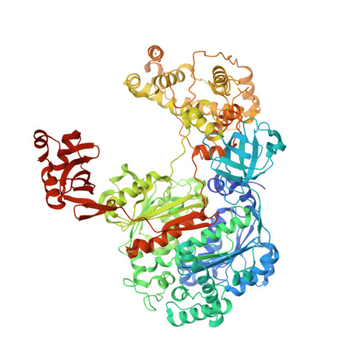
Structure of the ubiquitin-activating enzyme loaded with two ubiquitin molecules.

Schafer, A.Kuhn, M.Schindelin, H.

(2014) Acta Crystallogr D Biol Crystallogr 70: 1311-1320

  • DOI: https://doi.org/10.1107/S1399004714002910
  • Primary Citation Related Structures: 
    4NNJ

  • PubMed Abstract: 

    The activation of ubiquitin by the ubiquitin-activating enzyme Uba1 (E1) constitutes the first step in the covalent modification of target proteins with ubiquitin. This activation is a three-step process in which ubiquitin is adenylated at its C-terminal glycine, followed by the covalent attachment of ubiquitin to a catalytic cysteine residue of Uba1 and the subsequent adenylation of a second ubiquitin. Here, a ubiquitin E1 structure loaded with two ubiquitin molecules is presented for the first time. While one ubiquitin is bound in its adenylated form to the active adenylation domain of E1, the second ubiquitin represents the status after transfer and is covalently linked to the active-site cysteine. The covalently linked ubiquitin enables binding of the E2 enzyme without further modification of the ternary Uba1-ubiquitin2 arrangement. This doubly loaded E1 structure constitutes a missing link in the structural analysis of the ubiquitin-transfer cascade.


  • Organizational Affiliation
    • Structural Biology, Rudolf Virchow Center for Experimental Biomedicine, University of Würzburg, Josef-Schneider-Strasse 2, D-97080 Würzburg, Germany.

Macromolecule Content 

  • Total Structure Weight: 262.75 kDa 
  • Atom Count: 19,001 
  • Modeled Residue Count: 2,244 
  • Deposited Residue Count: 2,317 
  • Unique protein chains: 2

Macromolecules

Find similar proteins by:|  3D Structure
Entity ID: 1
MoleculeChains  Sequence LengthOrganismDetailsImage
Ubiquitin-activating enzyme E1 1
A, C
1,040Saccharomyces cerevisiae S288CMutation(s): 0 
Gene Names: UBA1YKL210W
EC: 6.2.1.45
UniProt
Find proteins for P22515 (Saccharomyces cerevisiae (strain ATCC 204508 / S288c))
Explore P22515 
Go to UniProtKB:  P22515
Entity Groups
Sequence Clusters30% Identity50% Identity70% Identity90% Identity95% Identity100% Identity
UniProt GroupP22515
Sequence Annotations
Expand
Reference Sequence
Find similar proteins by:|  3D Structure
Entity ID: 2
MoleculeChains  Sequence LengthOrganismDetailsImage
Uba1
B, D, E
79Saccharomyces cerevisiae S288CMutation(s): 0 
Gene Names: SCD2UBI4YLL039C
UniProt
Find proteins for P0CG63 (Saccharomyces cerevisiae (strain ATCC 204508 / S288c))
Explore P0CG63 
Go to UniProtKB:  P0CG63
Entity Groups
Sequence Clusters30% Identity50% Identity70% Identity90% Identity95% Identity100% Identity
UniProt GroupP0CG63
Sequence Annotations
Expand
Reference Sequence

Small Molecules

Ligands 3 Unique
IDChains Name / Formula / InChI Key2D Diagram3D Interactions
AMP

Query on AMP


Download:Ideal Coordinates CCD File
NA [auth D],
Y [auth B]
ADENOSINE MONOPHOSPHATE
C10 H14 N5 O7 P
UDMBCSSLTHHNCD-KQYNXXCUSA-N
SO4

Query on SO4


Download:Ideal Coordinates CCD File
F [auth A],
MA [auth C],
X [auth A],
Z [auth C]
SULFATE ION
O4 S
QAOWNCQODCNURD-UHFFFAOYSA-L
GOL

Query on GOL


Download:Ideal Coordinates CCD File
AA [auth C]
BA [auth C]
CA [auth C]
DA [auth C]
EA [auth C]
AA [auth C],
BA [auth C],
CA [auth C],
DA [auth C],
EA [auth C],
FA [auth C],
G [auth A],
GA [auth C],
H [auth A],
HA [auth C],
I [auth A],
IA [auth C],
J [auth A],
JA [auth C],
K [auth A],
KA [auth C],
L [auth A],
LA [auth C],
M [auth A],
N [auth A],
O [auth A],
P [auth A],
Q [auth A],
R [auth A],
S [auth A],
T [auth A],
U [auth A],
V [auth A],
W [auth A]
GLYCEROL
C3 H8 O3
PEDCQBHIVMGVHV-UHFFFAOYSA-N

Experimental Data & Validation

Experimental Data

  • Method: X-RAY DIFFRACTION
  • Resolution: 2.40 Å
  • R-Value Free:  0.201 (Depositor), 0.170 (DCC) 
  • R-Value Work:  0.164 (Depositor), 0.171 (DCC) 
  • R-Value Observed: 0.164 (Depositor) 
Space Group: P 2 21 21
Unit Cell:
Length ( Å )Angle ( ˚ )
a = 73.04α = 90
b = 195.651β = 90
c = 230.629γ = 90
Software Package:
Software NamePurpose
MxCuBEdata collection
EDNAdata collection
PHASERphasing
PHENIXrefinement
XDSdata reduction
SCALAdata scaling

Structure Validation

View Full Validation Report


Ligand Structure Quality Assessment 


Entry History 

Deposition Data

Revision History  (Full details and data files)

  • Version 1.0: 2014-05-07
    Type: Initial release
  • Version 1.1: 2014-09-24
    Changes: Database references
  • Version 1.2: 2023-09-20
    Changes: Data collection, Database references, Derived calculations, Refinement description
  • Version 1.3: 2024-10-16
    Changes: Structure summary