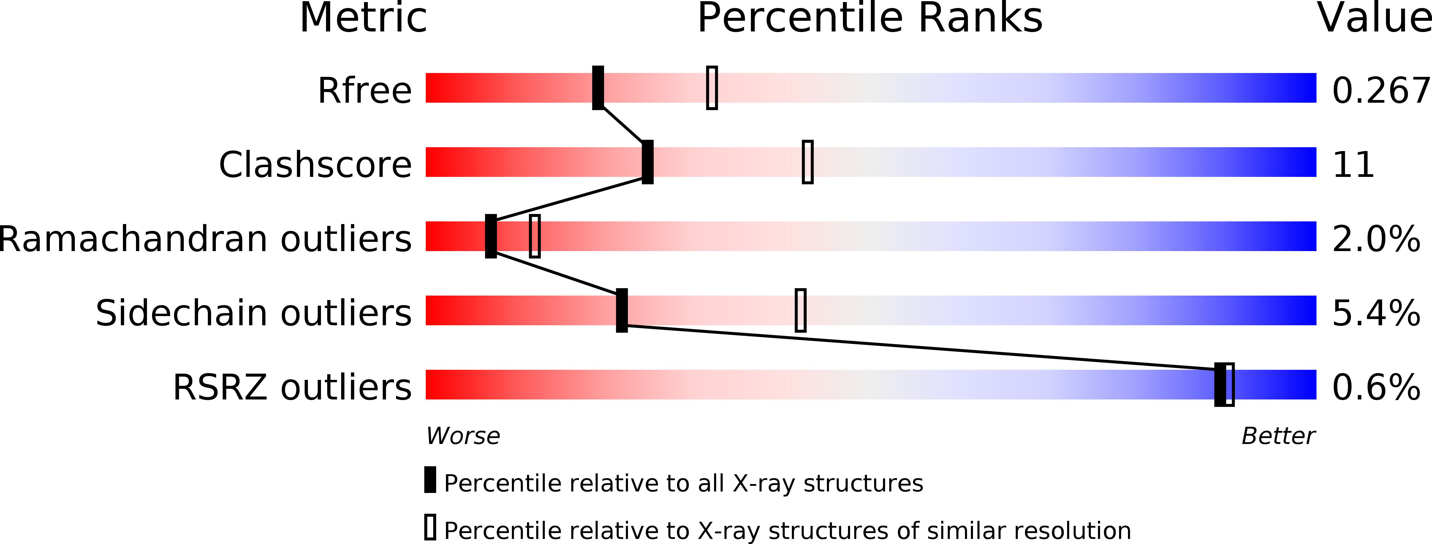
4NJE

Crystal structure of Pyrococcus furiosus L-asparaginase with ligand


Experimental Data Snapshot

  • Method: X-RAY DIFFRACTION
  • Resolution: 2.50 Å
  • R-Value Free: 0.270 
  • R-Value Work: 0.228 
  • R-Value Observed: 0.231 

Starting Model: experimental
View more details

wwPDB Validation   3D Report Full Report


This is version 1.2 of the entry. See complete history


Literature

Structural and functional insights into an archaeal L-asparaginase obtained through the linker-less assembly of constituent domains.

Tomar, R.Sharma, P.Srivastava, A.Bansal, S.Kundu, B.

(2014) Acta Crystallogr D Biol Crystallogr 70: 3187-3197

  • DOI: https://doi.org/10.1107/S1399004714023414
  • Primary Citation of Related Structures:  
    4NJE, 4Q0M, 4RA6, 4RA9

  • PubMed Abstract: 

    Covalent linkers bridging the domains of multidomain proteins are considered to be crucial for assembly and function. In this report, an exception in which the linker of a two-domain dimeric L-asparaginase from Pyrococcus furiosus (PfA) was found to be dispensable is presented. Domains of this enzyme assembled without the linker into a conjoined tetrameric form that exhibited higher activity than the parent enzyme. The global shape and quaternary structure of the conjoined PfA were also similar to the wild-type PfA, as observed by their solution scattering profiles and X-ray crystallographic data. Comparison of the crystal structures of substrate-bound and unbound enzymes revealed an altogether new active-site composition and mechanism of action. Thus, conjoined PfA is presented as a unique enzyme obtained through noncovalent, linker-less assembly of constituent domains that is stable enough to function efficiently at elevated temperatures.


  • Organizational Affiliation

    Kusuma School of Biological Sciences, Indian Institute of Technology Delhi, New Delhi, India.


Macromolecules
Find similar proteins by:  (by identity cutoff)  |  3D Structure
Entity ID: 1
MoleculeChains Sequence LengthOrganismDetailsImage
L-asparaginase204Pyrococcus furiosus DSM 3638Mutation(s): 0 
Gene Names: ph0066PF2047
EC: 3.5.1.1
UniProt
Find proteins for Q8TZE8 (Pyrococcus furiosus (strain ATCC 43587 / DSM 3638 / JCM 8422 / Vc1))
Explore Q8TZE8 
Go to UniProtKB:  Q8TZE8
Entity Groups  
Sequence Clusters30% Identity50% Identity70% Identity90% Identity95% Identity100% Identity
UniProt GroupQ8TZE8
Sequence Annotations
Expand
  • Reference Sequence
Find similar proteins by:  (by identity cutoff)  |  3D Structure
Entity ID: 2
MoleculeChains Sequence LengthOrganismDetailsImage
L-asparaginase147Pyrococcus furiosus DSM 3638Mutation(s): 0 
Gene Names: ph0066PF2047
EC: 3.5.1.1
UniProt
Find proteins for Q8TZE8 (Pyrococcus furiosus (strain ATCC 43587 / DSM 3638 / JCM 8422 / Vc1))
Explore Q8TZE8 
Go to UniProtKB:  Q8TZE8
Entity Groups  
Sequence Clusters30% Identity50% Identity70% Identity90% Identity95% Identity100% Identity
UniProt GroupQ8TZE8
Sequence Annotations
Expand
  • Reference Sequence
Small Molecules
Ligands 1 Unique
IDChains Name / Formula / InChI Key2D Diagram3D Interactions
ASP
Query on ASP

Download Ideal Coordinates CCD File 
C [auth A]ASPARTIC ACID
C4 H7 N O4
CKLJMWTZIZZHCS-REOHCLBHSA-N
Experimental Data & Validation

Experimental Data

  • Method: X-RAY DIFFRACTION
  • Resolution: 2.50 Å
  • R-Value Free: 0.270 
  • R-Value Work: 0.228 
  • R-Value Observed: 0.231 
  • Space Group: P 65 2 2
Unit Cell:
Length ( Å )Angle ( ˚ )
a = 91.59α = 90
b = 91.59β = 90
c = 188.77γ = 120
Software Package:
Software NamePurpose
MAR345dtbdata collection
PHASESphasing
PHENIXrefinement
MOSFLMdata reduction
SCALAdata scaling

Structure Validation

View Full Validation Report


Entry History 

Deposition Data

Revision History  (Full details and data files)

  • Version 1.0: 2014-12-10
    Type: Initial release
  • Version 1.1: 2020-01-01
    Changes: Database references
  • Version 1.2: 2023-11-08
    Changes: Data collection, Database references, Derived calculations, Refinement description