4NER | pdb_00004ner

Multicopper Oxidase CueO (data1)


Experimental Data Snapshot

  • Method: X-RAY DIFFRACTION
  • Resolution: 1.60 Å
  • R-Value Work: 
    0.168 (Depositor), 0.164 (DCC) 
  • R-Value Observed: 
    0.168 (Depositor) 

wwPDB Validation 3D Report Full Report

Validation slider image for 4NER

This is version 1.2 of the entry. See complete history

Literature

New insights into the catalytic active-site structure of multicopper oxidases.

Komori, H.Sugiyama, R.Kataoka, K.Miyazaki, K.Higuchi, Y.Sakurai, T.

(2014) Acta Crystallogr D Biol Crystallogr 70: 772-779

  • DOI: https://doi.org/10.1107/S1399004713033051
  • Primary Citation Related Structures: 
    4E9V, 4E9W, 4E9X, 4E9Y, 4NER

  • PubMed Abstract: 

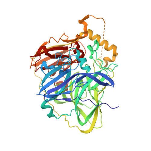
    Structural models determined by X-ray crystallography play a central role in understanding the catalytic mechanism of enzymes. However, X-ray radiation generates hydrated electrons that can cause significant damage to the active sites of metalloenzymes. In the present study, crystal structures of the multicopper oxidases (MCOs) CueO from Escherichia coli and laccase from a metagenome were determined. Diffraction data were obtained from a single crystal under low to high X-ray dose conditions. At low levels of X-ray exposure, unambiguous electron density for an O atom was observed inside the trinuclear copper centre (TNC) in both MCOs. The gradual reduction of copper by hydrated electrons monitored by measurement of the Cu K-edge X-ray absorption spectra led to the disappearance of the electron density for the O atom. In addition, the size of the copper triangle was enlarged by a two-step shift in the location of the type III coppers owing to reduction. Further, binding of O2 to the TNC after its full reduction was observed in the case of the laccase. Based on these novel structural findings, the diverse resting structures of the MCOs and their four-electron O2-reduction process are discussed.


  • Organizational Affiliation
    • Faculty of Education, Kagawa University, 1-1 Saiwai-cho, Takamatsu, Kagawa 760-8522, Japan.

Macromolecule Content 

  • Total Structure Weight: 53.83 kDa 
  • Atom Count: 4,215 
  • Modeled Residue Count: 476 
  • Deposited Residue Count: 489 
  • Unique protein chains: 1

Macromolecules

Find similar proteins by:|  3D Structure
Entity ID: 1
MoleculeChains  Sequence LengthOrganismDetailsImage
Blue copper oxidase CueO489Escherichia coli K-12Mutation(s): 0 
Gene Names: cueOyacKb0123JW0119
EC: 1.16.3.4
UniProt
Find proteins for P36649 (Escherichia coli (strain K12))
Explore P36649 
Go to UniProtKB:  P36649
Entity Groups
Sequence Clusters30% Identity50% Identity70% Identity90% Identity95% Identity100% Identity
UniProt GroupP36649
Sequence Annotations
Expand
Reference Sequence

Experimental Data & Validation

Experimental Data

  • Method: X-RAY DIFFRACTION
  • Resolution: 1.60 Å
  • R-Value Work:  0.168 (Depositor), 0.164 (DCC) 
  • R-Value Observed: 0.168 (Depositor) 
Space Group: P 1 21 1
Unit Cell:
Length ( Å )Angle ( ˚ )
a = 50.398α = 90
b = 90.721β = 102.72
c = 53.37γ = 90
Software Package:
Software NamePurpose
SHELXrefinement
PDB_EXTRACTdata extraction
SHELXLrefinement

Structure Validation

View Full Validation Report


Entry History 

Deposition Data

Revision History  (Full details and data files)

  • Version 1.0: 2014-03-12
    Type: Initial release
  • Version 1.1: 2019-12-25
    Changes: Database references
  • Version 1.2: 2024-03-20
    Changes: Data collection, Database references, Derived calculations