4ND2 | pdb_00004nd2

Crystal structure of the lactate dehydrogenase from cryptosporidium parvum complexed with substrate (pyruvic acid) and cofactor analog (3-acetylpyridine adenine dinucleotide)


Experimental Data Snapshot

  • Method: X-RAY DIFFRACTION
  • Resolution: 2.00 Å
  • R-Value Free: 
    0.208 (Depositor), 0.216 (DCC) 
  • R-Value Work: 
    0.191 (Depositor), 0.199 (DCC) 
  • R-Value Observed: 
    0.193 (Depositor) 

Starting Model: experimental
View more details

wwPDB Validation 3D Report Full Report

Validation slider image for 4ND2

Ligand Structure Quality Assessment 


This is version 2.1 of the entry. See complete history

Literature

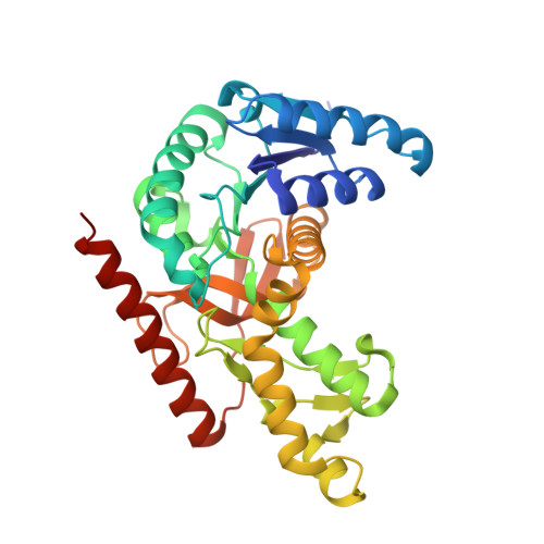
Biochemical and structural characterization of Cryptosporidium parvum Lactate dehydrogenase.

Cook, W.J.Senkovich, O.Hernandez, A.Speed, H.Chattopadhyay, D.

(2014) Int J Biol Macromol 74C: 608-619

  • DOI: https://doi.org/10.1016/j.ijbiomac.2014.12.019
  • Primary Citation Related Structures: 
    4ND1, 4ND2, 4ND3, 4ND4, 4ND5

  • PubMed Abstract: 

    The protozoan parasite Cryptosporidium parvum causes waterborne diseases worldwide. There is no effective therapy for C. parvum infection. The parasite depends mainly on glycolysis for energy production. Lactate dehydrogenase is a major regulator of glycolysis. This paper describes the biochemical characterization of C. parvum lactate dehydrogenase and high resolution crystal structures of the apo-enzyme and four ternary complexes. The ternary complexes capture the enzyme bound to NAD/NADH or its 3-acetylpyridine analog in the cofactor binding pocket, while the substrate binding site is occupied by one of the following ligands: lactate, pyruvate or oxamate. The results reveal distinctive features of the parasitic enzyme. For example, C. parvum lactate dehydrogenase prefers the acetylpyridine analog of NADH as a cofactor. Moreover, it is slightly less sensitive to gossypol inhibition compared with mammalian lactate dehydrogenases and not inhibited by excess pyruvate. The active site loop and the antigenic loop in C. parvum lactate dehydrogenase are considerably different from those in the human counterpart. Structural features and enzymatic properties of C. parvum lactate dehydrogenase are similar to enzymes from related parasites. Structural comparison with malate dehydrogenase supports a common ancestry for the two genes.


  • Organizational Affiliation
    • Department of Pathology, University of Alabama at Birmingham, Birmingham, AL 35294, United States.

Macromolecule Content 

  • Total Structure Weight: 69.55 kDa 
  • Atom Count: 5,241 
  • Modeled Residue Count: 634 
  • Deposited Residue Count: 642 
  • Unique protein chains: 1

Macromolecules

Find similar proteins by:|  3D Structure
Entity ID: 1
MoleculeChains  Sequence LengthOrganismDetailsImage
Lactate dehydrogenase, adjacent gene encodes predicted malate dehydrogenase
A, B
321Cryptosporidium parvum Iowa IIMutation(s): 0 
Gene Names: cgd7_480LDH1
EC: 1.1.1.27
UniProt
Find proteins for Q5CYZ2 (Cryptosporidium parvum (strain Iowa II))
Explore Q5CYZ2 
Go to UniProtKB:  Q5CYZ2
Entity Groups
Sequence Clusters30% Identity50% Identity70% Identity90% Identity95% Identity100% Identity
UniProt GroupQ5CYZ2
Sequence Annotations
Expand
Reference Sequence

Small Molecules

Ligands 3 Unique
IDChains Name / Formula / InChI Key2D Diagram3D Interactions
A3D

Query on A3D


Download:Ideal Coordinates CCD File
C [auth A],
G [auth B]
3-ACETYLPYRIDINE ADENINE DINUCLEOTIDE
C22 H28 N6 O14 P2
KPVQNXLUPNWQHM-RBEMOOQDSA-N
GOL

Query on GOL


Download:Ideal Coordinates CCD File
E [auth A],
F [auth A],
I [auth B]
GLYCEROL
C3 H8 O3
PEDCQBHIVMGVHV-UHFFFAOYSA-N
PYR

Query on PYR


Download:Ideal Coordinates CCD File
D [auth A],
H [auth B]
PYRUVIC ACID
C3 H4 O3
LCTONWCANYUPML-UHFFFAOYSA-N

Experimental Data & Validation

Experimental Data

  • Method: X-RAY DIFFRACTION
  • Resolution: 2.00 Å
  • R-Value Free:  0.208 (Depositor), 0.216 (DCC) 
  • R-Value Work:  0.191 (Depositor), 0.199 (DCC) 
  • R-Value Observed: 0.193 (Depositor) 
Space Group: P 32 2 1
Unit Cell:
Length ( Å )Angle ( ˚ )
a = 95.83α = 90
b = 95.83β = 90
c = 185.64γ = 120
Software Package:
Software NamePurpose
REFMACrefinement
CNSrefinement
DENZOdata reduction
SCALEPACKdata scaling
CNSphasing

Structure Validation

View Full Validation Report


Ligand Structure Quality Assessment 


Entry History 

Deposition Data

Revision History  (Full details and data files)

  • Version 1.0: 2014-12-17
    Type: Initial release
  • Version 1.1: 2015-03-11
    Changes: Database references
  • Version 1.2: 2019-07-17
    Changes: Data collection, Derived calculations, Refinement description
  • Version 1.3: 2023-09-20
    Changes: Data collection, Database references, Derived calculations, Refinement description
  • Version 2.0: 2023-11-15
    Changes: Atomic model, Data collection
  • Version 2.1: 2024-10-16
    Changes: Structure summary