4N7Q | pdb_00004n7q

Crystal structure of eukaryotic THIC from A. thaliana


Experimental Data Snapshot

  • Method: X-RAY DIFFRACTION
  • Resolution: 1.60 Å
  • R-Value Free: 
    0.176 (Depositor), 0.176 (DCC) 
  • R-Value Work: 
    0.152 (Depositor), 0.152 (DCC) 
  • R-Value Observed: 
    0.154 (Depositor) 

Starting Model: experimental
View more details

wwPDB Validation 3D Report Full Report

Validation slider image for 4N7Q

This is version 1.2 of the entry. See complete history

Literature

High-resolution crystal structure of the eukaryotic HMP-P synthase (THIC) from Arabidopsis thaliana.

Coquille, S.Roux, C.Mehta, A.Begley, T.P.Fitzpatrick, T.B.Thore, S.

(2013) J Struct Biol 184: 438-444

  • DOI: https://doi.org/10.1016/j.jsb.2013.10.005
  • Primary Citation Related Structures: 
    4N7Q

  • PubMed Abstract: 

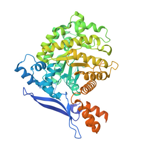
    Vitamin B₁ is an essential compound in all organisms acting as a cofactor in key metabolic reactions. It is formed by the condensation of two independently biosynthesized molecules referred to as the pyrimidine and thiazole moieties. In bacteria and plants, the biosynthesis of the pyrimidine moiety, 4-amino-5-hydroxymethyl-2-methylpyrimidine phosphate (HMP-P), requires a single enzyme, THIC (HMP-P synthase). The enzyme uses an iron-sulfur cluster as well as a 5'-deoxyadenosyl radical as cofactors to rearrange the 5-amino-imidazole ribonucleotide (AIR) substrate to the pyrimidine ring. So far, the only structure reported is the one from the bacteria Caulobacter crescentus. In an attempt to structurally characterize an eukaryotic HMP-P synthase, we have determined the high-resolution crystal structure of THIC from Arabidopsis thaliana at 1.6 Å. The structure is highly similar to its bacterial counterpart although several loop regions show significant differences with potential implications for the enzymatic properties. Furthermore, we have found a metal ion with octahedral coordination at the same location as a zinc ion in the bacterial enzyme. Our high-resolution atomic model shows a metal ion with multiple coordinated water molecules in the close vicinity of the substrate binding sites and is an important step toward the full characterization of the chemical rearrangement occurring during HMP-P biosynthesis.


  • Organizational Affiliation
    • Department of Molecular Biology, University of Geneva, Geneva 1211, Switzerland. Electronic address: Sandrine.coquille@unige.ch.

Macromolecule Content 

  • Total Structure Weight: 65.96 kDa 
  • Atom Count: 4,541 
  • Modeled Residue Count: 472 
  • Deposited Residue Count: 582 
  • Unique protein chains: 1

Macromolecules

Find similar proteins by:|  3D Structure
Entity ID: 1
MoleculeChains  Sequence LengthOrganismDetailsImage
Phosphomethylpyrimidine synthase, chloroplastic582Arabidopsis thalianaMutation(s): 0 
Gene Names: THICPYAt2g29630T27A16.27
EC: 4.1.99.17
UniProt
Find proteins for O82392 (Arabidopsis thaliana)
Explore O82392 
Go to UniProtKB:  O82392
Entity Groups
Sequence Clusters30% Identity50% Identity70% Identity90% Identity95% Identity100% Identity
UniProt GroupO82392
Sequence Annotations
Expand
Reference Sequence

Experimental Data & Validation

Experimental Data

  • Method: X-RAY DIFFRACTION
  • Resolution: 1.60 Å
  • R-Value Free:  0.176 (Depositor), 0.176 (DCC) 
  • R-Value Work:  0.152 (Depositor), 0.152 (DCC) 
  • R-Value Observed: 0.154 (Depositor) 
Space Group: P 32 2 1
Unit Cell:
Length ( Å )Angle ( ˚ )
a = 107α = 90
b = 107β = 90
c = 88.3γ = 120
Software Package:
Software NamePurpose
XDSdata scaling
CCP4model building
PHENIXrefinement
XDSdata reduction
CCP4phasing

Structure Validation

View Full Validation Report


Entry History 

Deposition Data

Revision History  (Full details and data files)

  • Version 1.0: 2013-11-13
    Type: Initial release
  • Version 1.1: 2013-12-18
    Changes: Database references
  • Version 1.2: 2023-09-20
    Changes: Data collection, Database references, Derived calculations, Refinement description