4N6M

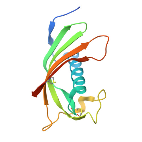
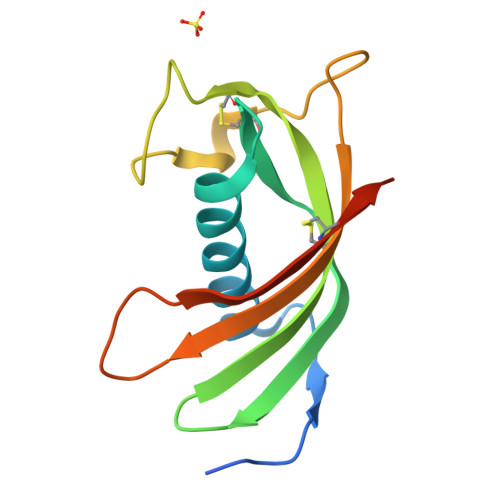
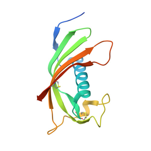
Crystal structure of human cystatin E/M produced in LEXSY


Experimental Data Snapshot

  • Method: X-RAY DIFFRACTION
  • Resolution: 2.90 Å
  • R-Value Free: 0.283 
  • R-Value Work: 0.261 
  • R-Value Observed: 0.261 

Starting Model: experimental
View more details

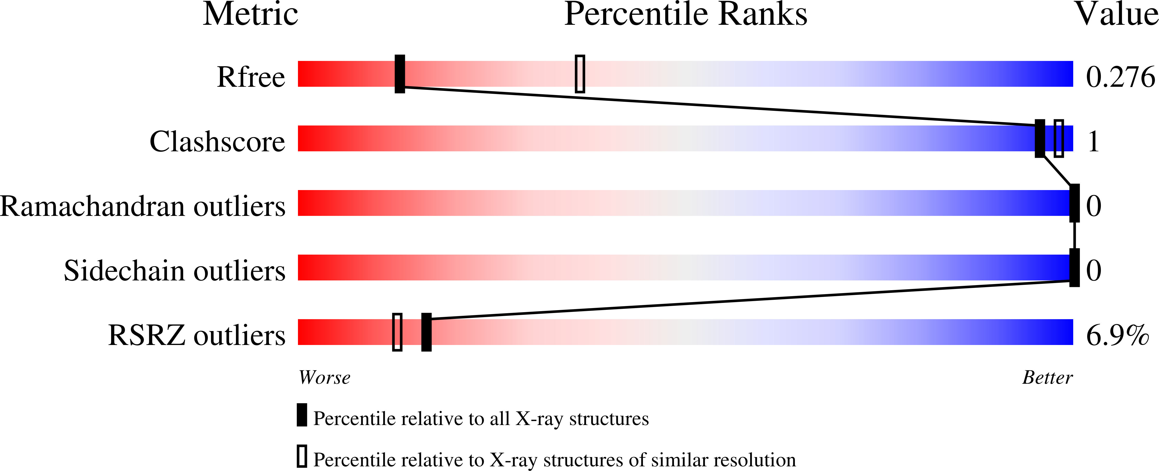
wwPDB Validation   3D Report Full Report


This is version 1.3 of the entry. See complete history


Literature

Structure and mechanism of an aspartimide-dependent Peptide ligase in human legumain.

Dall, E.Fegg, J.C.Briza, P.Brandstetter, H.

(2015) Angew Chem Int Ed Engl 54: 2917-2921

  • DOI: https://doi.org/10.1002/anie.201409135
  • Primary Citation of Related Structures:  
    4N6L, 4N6M, 4N6N, 4N6O

  • PubMed Abstract: 

    Peptide ligases expand the repertoire of genetically encoded protein architectures by synthesizing new peptide bonds, energetically driven by ATP or NTPs. Here, we report the discovery of a genuine ligase activity in human legumain (AEP) which has important roles in immunity and tumor progression that were believed to be due to its established cysteine protease activity. Defying dogma, the ligase reaction is independent of the catalytic cysteine but exploits an endogenous energy reservoir that results from the conversion of a conserved aspartate to a metastable aspartimide. Legumain's dual protease-ligase activities are pH- and thus localization controlled, dominating at acidic and neutral pH, respectively. Their relevance includes reversible on-off switching of cystatin inhibitors and enzyme (in)activation, and may affect the generation of three-dimensional MHC epitopes. The aspartate-aspartimide (succinimide) pair represents a new paradigm of coupling endergonic reactions in ATP-scarce environments.


  • Organizational Affiliation

    Department of Molecular Biology, University of Salzburg, 5020 Salzburg (Austria).


Macromolecules
Find similar proteins by:  (by identity cutoff)  |  3D Structure
Entity ID: 1
MoleculeChains Sequence LengthOrganismDetailsImage
Cystatin-M
A, B
132Homo sapiensMutation(s): 0 
Gene Names: CST6
UniProt & NIH Common Fund Data Resources
Find proteins for Q15828 (Homo sapiens)
Explore Q15828 
Go to UniProtKB:  Q15828
PHAROS:  Q15828
GTEx:  ENSG00000175315 
Entity Groups  
Sequence Clusters30% Identity50% Identity70% Identity90% Identity95% Identity100% Identity
UniProt GroupQ15828
Sequence Annotations
Expand
  • Reference Sequence
Small Molecules
Ligands 1 Unique
IDChains Name / Formula / InChI Key2D Diagram3D Interactions
SO4
Query on SO4

Download Ideal Coordinates CCD File 
C [auth B]SULFATE ION
O4 S
QAOWNCQODCNURD-UHFFFAOYSA-L
Experimental Data & Validation

Experimental Data

  • Method: X-RAY DIFFRACTION
  • Resolution: 2.90 Å
  • R-Value Free: 0.283 
  • R-Value Work: 0.261 
  • R-Value Observed: 0.261 
  • Space Group: P 1 21 1
Unit Cell:
Length ( Å )Angle ( ˚ )
a = 39.71α = 90
b = 82.63β = 112.81
c = 43.49γ = 90
Software Package:
Software NamePurpose
MxCuBEdata collection
PHASERphasing
REFMACrefinement
iMOSFLMdata reduction
SCALAdata scaling

Structure Validation

View Full Validation Report


Entry History 

Deposition Data

Revision History  (Full details and data files)

  • Version 1.0: 2015-02-18
    Type: Initial release
  • Version 1.1: 2015-03-11
    Changes: Database references
  • Version 1.2: 2023-09-20
    Changes: Data collection, Database references, Derived calculations, Refinement description
  • Version 1.3: 2024-11-20
    Changes: Structure summary