4N4W | pdb_00004n4w

Structure of the human smoothened receptor in complex with SANT-1.


Experimental Data Snapshot

  • Method: X-RAY DIFFRACTION
  • Resolution: 2.80 Å
  • R-Value Free: 
    0.253 (Depositor), 0.254 (DCC) 
  • R-Value Work: 
    0.204 (Depositor) 
  • R-Value Observed: 
    0.207 (Depositor) 

Starting Model: experimental
View more details

wwPDB Validation 3D Report Full Report

Validation slider image for 4N4W

Ligand Structure Quality Assessment 


This is version 2.2 of the entry. See complete history

Literature

Structural basis for Smoothened receptor modulation and chemoresistance to anticancer drugs.

Wang, C.Wu, H.Evron, T.Vardy, E.Han, G.W.Huang, X.P.Hufeisen, S.J.Mangano, T.J.Urban, D.J.Katritch, V.Cherezov, V.Caron, M.G.Roth, B.L.Stevens, R.C.

(2014) Nat Commun 5: 4355-4355

  • DOI: https://doi.org/10.1038/ncomms5355
  • Primary Citation Related Structures: 
    4N4W, 4QIM, 4QIN

  • PubMed Abstract: 

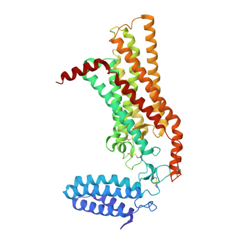
    The Smoothened receptor (SMO) mediates signal transduction in the hedgehog pathway, which is implicated in normal development and carcinogenesis. SMO antagonists can suppress the growth of some tumours; however, mutations at SMO have been found to abolish their antitumour effects, a phenomenon known as chemoresistance. Here we report three crystal structures of human SMO bound to the antagonists SANT1 and Anta XV, and the agonist, SAG1.5, at 2.6-2.8 Å resolution. The long and narrow cavity in the transmembrane domain of SMO harbours multiple ligand binding sites, where SANT1 binds at a deeper site as compared with other ligands. Distinct interactions at D473(6.54f) elucidated the structural basis for the differential effects of chemoresistance mutations on SMO antagonists. The agonist SAG1.5 induces a conformational rearrangement of the binding pocket residues, which could contribute to SMO activation. Collectively, these studies reveal the structural basis for the modulation of SMO by small molecules.


  • Organizational Affiliation
    • Department of Integrative Structural and Computational Biology, The Scripps Research Institute, 10550 North Torrey Pines Road, La Jolla, California 92037, USA.

Macromolecule Content 

  • Total Structure Weight: 55.39 kDa 
  • Atom Count: 3,707 
  • Modeled Residue Count: 457 
  • Deposited Residue Count: 475 
  • Unique protein chains: 1

Macromolecules

Find similar proteins by:|  3D Structure
Entity ID: 1
MoleculeChains  Sequence LengthOrganismDetailsImage
Cytochrome b(562),Smoothened homolog475Shigella flexneri 5 str. 8401Homo sapiens
This entity is chimeric
Mutation(s): 3 
Gene Names: cybCSFV_4255SMOSMOH
Membrane Entity: Yes 
UniProt & NIH Common Fund Data Resources
Find proteins for Q0SXH8 (Shigella flexneri serotype 5b (strain 8401))
Explore Q0SXH8 
Go to UniProtKB:  Q0SXH8
Find proteins for Q99835 (Homo sapiens)
Explore Q99835 
Go to UniProtKB:  Q99835
PHAROS:  Q99835
GTEx:  ENSG00000128602 
Entity Groups
Sequence Clusters30% Identity50% Identity70% Identity90% Identity95% Identity100% Identity
UniProt GroupsQ99835Q0SXH8
Sequence Annotations
Expand
Reference Sequence

Oligosaccharides

Help  
Entity ID: 2
MoleculeChains Length2D Diagram GlycosylationD Interactions
alpha-D-mannopyranose-(1-3)-beta-D-mannopyranose-(1-4)-2-acetamido-2-deoxy-beta-D-glucopyranose-(1-4)-2-acetamido-2-deoxy-beta-D-glucopyranose
B
4N/A
Glycosylation Resources
GlyTouCan: G81315DD
GlyCosmos: G81315DD
GlyGen: G81315DD

Small Molecules

Ligands 5 Unique
IDChains Name / Formula / InChI Key2D Diagram3D Interactions
SNT

Query on SNT


Download:Ideal Coordinates CCD File
C [auth A](E)-N-(4-benzylpiperazin-1-yl)-1-(3,5-dimethyl-1-phenyl-1H-pyrazol-4-yl)methanimine
C23 H27 N5
FOORCIAZMIWALX-JJIBRWJFSA-N
OLC

Query on OLC


Download:Ideal Coordinates CCD File
F [auth A],
G [auth A]
(2R)-2,3-dihydroxypropyl (9Z)-octadec-9-enoate
C21 H40 O4
RZRNAYUHWVFMIP-GDCKJWNLSA-N
PG4

Query on PG4


Download:Ideal Coordinates CCD File
I [auth A]TETRAETHYLENE GLYCOL
C8 H18 O5
UWHCKJMYHZGTIT-UHFFFAOYSA-N
PEG

Query on PEG


Download:Ideal Coordinates CCD File
H [auth A]DI(HYDROXYETHYL)ETHER
C4 H10 O3
MTHSVFCYNBDYFN-UHFFFAOYSA-N
ZN

Query on ZN


Download:Ideal Coordinates CCD File
D [auth A],
E [auth A]
ZINC ION
Zn
PTFCDOFLOPIGGS-UHFFFAOYSA-N

Experimental Data & Validation

Experimental Data

  • Method: X-RAY DIFFRACTION
  • Resolution: 2.80 Å
  • R-Value Free:  0.253 (Depositor), 0.254 (DCC) 
  • R-Value Work:  0.204 (Depositor) 
  • R-Value Observed: 0.207 (Depositor) 
Space Group: C 2 2 21
Unit Cell:
Length ( Å )Angle ( ˚ )
a = 84.64α = 90
b = 110.88β = 90
c = 145.21γ = 90
Software Package:
Software NamePurpose
Blu-Icedata collection
PHASERphasing
BUSTERrefinement
HKL-2000data reduction
HKL-2000data scaling

Structure Validation

View Full Validation Report


Ligand Structure Quality Assessment 


Entry History 

Deposition Data

Revision History  (Full details and data files)

  • Version 1.0: 2014-01-22
    Type: Initial release
  • Version 1.1: 2014-07-30
    Changes: Database references
  • Version 1.2: 2017-06-07
    Changes: Database references, Structure summary
  • Version 1.3: 2017-11-15
    Changes: Refinement description
  • Version 2.0: 2020-07-29
    Type: Remediation
    Reason: Carbohydrate remediation
    Changes: Advisory, Atomic model, Data collection, Derived calculations, Structure summary
  • Version 2.1: 2023-09-20
    Changes: Data collection, Database references, Refinement description, Structure summary
  • Version 2.2: 2024-11-20
    Changes: Structure summary