4MZT | pdb_00004mzt

MazF from S. aureus crystal form II, C2221, 2.3 A


Experimental Data Snapshot

  • Method: X-RAY DIFFRACTION
  • Resolution: 2.30 Å
  • R-Value Free: 
    0.248 (Depositor), 0.239 (DCC) 
  • R-Value Work: 
    0.196 (Depositor), 0.192 (DCC) 
  • R-Value Observed: 
    0.198 (Depositor) 

Starting Model: experimental
View more details

wwPDB Validation 3D Report Full Report

Validation slider image for 4MZT

This is version 1.2 of the entry. See complete history

Literature

Structural and biophysical characterization of Staphylococcus aureus SaMazF shows conservation of functional dynamics.

Zorzini, V.Buts, L.Sleutel, M.Garcia-Pino, A.Talavera, A.Haesaerts, S.Greve, H.D.Cheung, A.van Nuland, N.A.Loris, R.

(2014) Nucleic Acids Res 42: 6709-6725

  • DOI: https://doi.org/10.1093/nar/gku266
  • Primary Citation Related Structures: 
    4MZM, 4MZP, 4MZT

  • PubMed Abstract: 

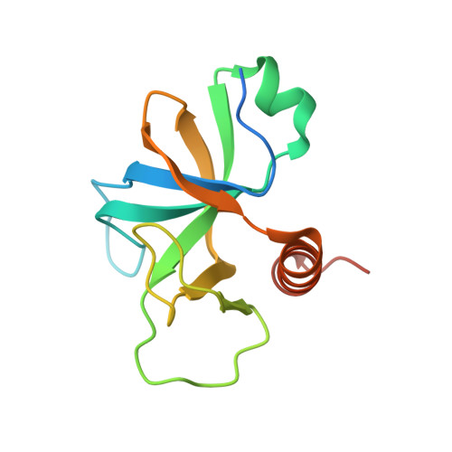
    The Staphylococcus aureus genome contains three toxin-antitoxin modules, including one mazEF module, SamazEF. Using an on-column separation protocol we are able to obtain large amounts of wild-type SaMazF toxin. The protein is well-folded and highly resistant against thermal unfolding but aggregates at elevated temperatures. Crystallographic and nuclear magnetic resonance (NMR) solution studies show a well-defined dimer. Differences in structure and dynamics between the X-ray and NMR structural ensembles are found in three loop regions, two of which undergo motions that are of functional relevance. The same segments also show functionally relevant dynamics in the distantly related CcdB family despite divergence of function. NMR chemical shift mapping and analysis of residue conservation in the MazF family suggests a conserved mode for the inhibition of MazF by MazE.


  • Organizational Affiliation
    • Structural Biology Brussels, Vrije Universiteit Brussel, Pleinlaan 2, 1050 Brussels, Belgium Molecular Recognition Unit, Department of Structural Biology, VIB, Pleinlaan 2, 1050 Brussels, Belgium.

Macromolecule Content 

  • Total Structure Weight: 29.9 kDa 
  • Atom Count: 1,743 
  • Modeled Residue Count: 227 
  • Deposited Residue Count: 266 
  • Unique protein chains: 1

Macromolecules

Find similar proteins by:|  3D Structure
Entity ID: 1
MoleculeChains  Sequence LengthOrganismDetailsImage
MazF mRNA interferase
A, B
133Staphylococcus aureus subsp. aureus N315Mutation(s): 0 
Gene Names: mazFSA1873
EC: 3.1
UniProt
Find proteins for Q7A4G9 (Staphylococcus aureus (strain N315))
Explore Q7A4G9 
Go to UniProtKB:  Q7A4G9
Entity Groups
Sequence Clusters30% Identity50% Identity70% Identity90% Identity95% Identity100% Identity
UniProt GroupQ7A4G9
Sequence Annotations
Expand
Reference Sequence

Experimental Data & Validation

Experimental Data

  • Method: X-RAY DIFFRACTION
  • Resolution: 2.30 Å
  • R-Value Free:  0.248 (Depositor), 0.239 (DCC) 
  • R-Value Work:  0.196 (Depositor), 0.192 (DCC) 
  • R-Value Observed: 0.198 (Depositor) 
Space Group: C 2 2 21
Unit Cell:
Length ( Å )Angle ( ˚ )
a = 72.588α = 90
b = 92.007β = 90
c = 71.52γ = 90
Software Package:
Software NamePurpose
ADSCdata collection
CCP4model building
PHENIXrefinement
HKL-2000data reduction
HKL-2000data scaling
CCP4phasing

Structure Validation

View Full Validation Report


Entry History 

Deposition Data

Revision History  (Full details and data files)

  • Version 1.0: 2014-05-21
    Type: Initial release
  • Version 1.1: 2014-06-25
    Changes: Database references
  • Version 1.2: 2023-09-20
    Changes: Data collection, Database references, Refinement description