4MRT

Structure of the Phosphopantetheine Transferase Sfp in Complex with Coenzyme A and a Peptidyl Carrier Protein


Experimental Data Snapshot

  • Method: X-RAY DIFFRACTION
  • Resolution: 2.00 Å
  • R-Value Free: 0.232 
  • R-Value Work: 0.188 
  • R-Value Observed: 0.191 

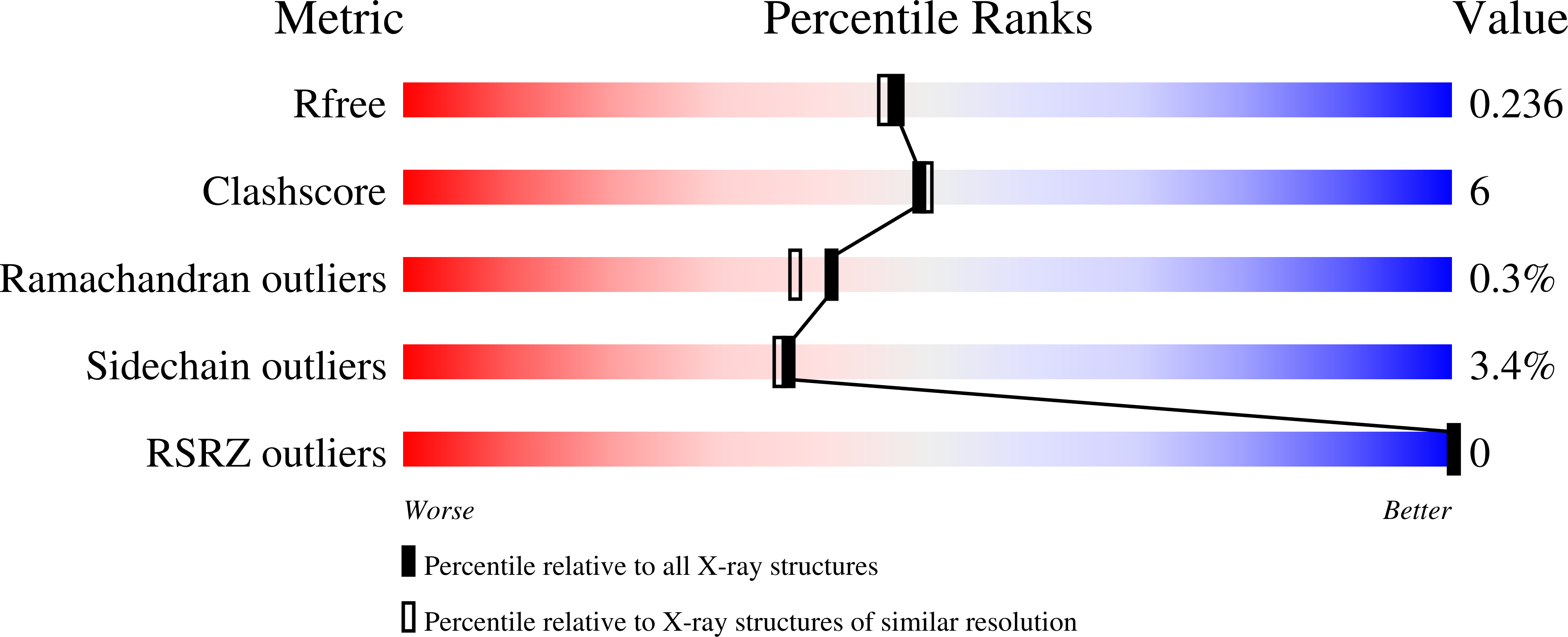
wwPDB Validation   3D Report Full Report


Ligand Structure Quality Assessment 


This is version 1.2 of the entry. See complete history


Literature

Crystal Structure of a PCP/Sfp Complex Reveals the Structural Basis for Carrier Protein Posttranslational Modification.

Tufar, P.Rahighi, S.Kraas, F.I.Kirchner, D.K.Lohr, F.Henrich, E.Kopke, J.Dikic, I.Guntert, P.Marahiel, M.A.Dotsch, V.

(2014) Chem Biol 21: 552-562

  • DOI: https://doi.org/10.1016/j.chembiol.2014.02.014
  • Primary Citation of Related Structures:  
    2MD9, 4MRT

  • PubMed Abstract: 

    Phosphopantetheine transferases represent a class of enzymes found throughout all forms of life. From a structural point of view, they are subdivided into three groups, with transferases from group II being the most widespread. They are required for the posttranslational modification of carrier proteins involved in diverse metabolic pathways. We determined the crystal structure of the group II phosphopantetheine transferase Sfp from Bacillus in complex with a substrate carrier protein in the presence of coenzyme A and magnesium, and observed two protein-protein interaction sites. Mutational analysis showed that only the hydrophobic contacts between the carrier protein's second helix and the C-terminal domain of Sfp are essential for their productive interaction. Comparison with a similar structure of a complex of human proteins suggests that the mode of interaction is highly conserved in all domains of life.


  • Organizational Affiliation

    Institute of Biophysical Chemistry and Center for Biomolecular Magnetic Resonance, Goethe University Frankfurt/Main, Max-von-Laue-Strasse 9, 60438 Frankfurt, Germany; Buchmann Institute for Molecular Life Sciences, Goethe University Frankfurt/Main, Max-von-Laue-Strasse 15, 60438 Frankfurt, Germany.


Macromolecules
Find similar proteins by:  (by identity cutoff)  |  3D Structure
Entity ID: 1
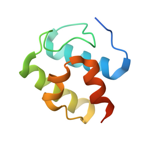
MoleculeChains Sequence LengthOrganismDetailsImage
Tyrocidine synthase 3A [auth C]90Brevibacillus parabrevisMutation(s): 1 
Gene Names: tycC
UniProt
Find proteins for O30409 (Brevibacillus parabrevis)
Explore O30409 
Go to UniProtKB:  O30409
Entity Groups  
Sequence Clusters30% Identity50% Identity70% Identity90% Identity95% Identity100% Identity
UniProt GroupO30409
Sequence Annotations
Expand
  • Reference Sequence
Find similar proteins by:  (by identity cutoff)  |  3D Structure
Entity ID: 2
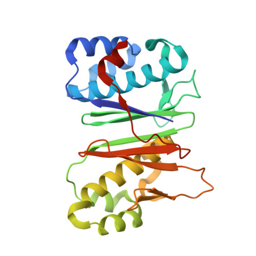
MoleculeChains Sequence LengthOrganismDetailsImage
4'-phosphopantetheinyl transferase sfpB [auth A]232Bacillus subtilis subsp. subtilis str. 168Mutation(s): 2 
Gene Names: BSU03570lpa-8sfp
EC: 2.7.8 (PDB Primary Data), 2.7.8.7 (UniProt)
UniProt
Find proteins for P39135 (Bacillus subtilis (strain 168))
Explore P39135 
Go to UniProtKB:  P39135
Entity Groups  
Sequence Clusters30% Identity50% Identity70% Identity90% Identity95% Identity100% Identity
UniProt GroupP39135
Sequence Annotations
Expand
  • Reference Sequence
Experimental Data & Validation

Experimental Data

  • Method: X-RAY DIFFRACTION
  • Resolution: 2.00 Å
  • R-Value Free: 0.232 
  • R-Value Work: 0.188 
  • R-Value Observed: 0.191 
  • Space Group: C 1 2 1
Unit Cell:
Length ( Å )Angle ( ˚ )
a = 160.66α = 90
b = 39.09β = 107.03
c = 53.31γ = 90
Software Package:
Software NamePurpose
MOSFLMdata reduction
SCALAdata scaling
MOLREPphasing
REFMACrefinement
PDB_EXTRACTdata extraction
go.comdata collection

Structure Validation

View Full Validation Report


Ligand Structure Quality Assessment 


Entry History 

Deposition Data

Revision History  (Full details and data files)

  • Version 1.0: 2014-04-23
    Type: Initial release
  • Version 1.1: 2014-05-14
    Changes: Database references
  • Version 1.2: 2024-02-28
    Changes: Data collection, Database references, Derived calculations