4MN8 | pdb_00004mn8

Crystal structure of flg22 in complex with the FLS2 and BAK1 ectodomains


Experimental Data Snapshot

  • Method: X-RAY DIFFRACTION
  • Resolution: 3.06 Å
  • R-Value Free: 
    0.275 (Depositor), 0.279 (DCC) 
  • R-Value Work: 
    0.224 (Depositor), 0.231 (DCC) 
  • R-Value Observed: 
    0.227 (Depositor) 

Starting Model: experimental
View more details

wwPDB Validation 3D Report Full Report

Validation slider image for 4MN8

Ligand Structure Quality Assessment 


This is version 2.2 of the entry. See complete history

Literature

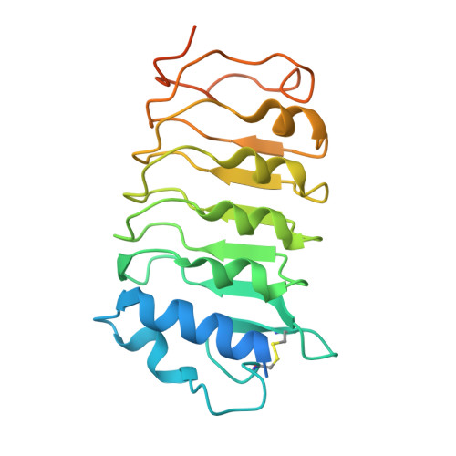
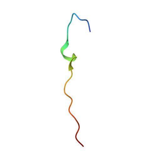
Structural basis for flg22-induced activation of the Arabidopsis FLS2-BAK1 immune complex.

Sun, Y.Li, L.Macho, A.P.Han, Z.Hu, Z.Zipfel, C.Zhou, J.M.Chai, J.

(2013) Science 342: 624-628

  • DOI: https://doi.org/10.1126/science.1243825
  • Primary Citation Related Structures: 
    4MN8, 4MNA

  • PubMed Abstract: 

    Flagellin perception in Arabidopsis is through recognition of its highly conserved N-terminal epitope (flg22) by flagellin-sensitive 2 (FLS2). Flg22 binding induces FLS2 heteromerization with BRASSINOSTEROID INSENSITIVE 1-associated kinase 1 (BAK1) and their reciprocal activation followed by plant immunity. Here, we report the crystal structure of FLS2 and BAK1 ectodomains complexed with flg22 at 3.06 angstroms. A conserved and a nonconserved site from the inner surface of the FLS2 solenoid recognize the C- and N-terminal segment of flg22, respectively, without oligomerization or conformational changes in the FLS2 ectodomain. Besides directly interacting with FLS2, BAK1 acts as a co-receptor by recognizing the C terminus of the FLS2-bound flg22. Our data reveal the molecular mechanisms underlying FLS2-BAK1 complex recognition of flg22 and provide insight into the immune receptor complex activation.


  • Organizational Affiliation
    • School of Life Sciences, Tsinghua University, Beijing 100084, China, and Tsinghua-Peking Center for Life Sciences, Beijing 100084, China.

Macromolecule Content 

  • Total Structure Weight: 116.38 kDa 
  • Atom Count: 7,487 
  • Modeled Residue Count: 955 
  • Deposited Residue Count: 1,030 
  • Unique protein chains: 3

Macromolecules

Find similar proteins by:|  3D Structure
Entity ID: 1
MoleculeChains  Sequence LengthOrganismDetailsImage
LRR receptor-like serine/threonine-protein kinase FLS2782Arabidopsis thalianaMutation(s): 0 
Gene Names: FLS2At5g46330MPL12.13MPL12.8
EC: 2.7.11.1
UniProt
Find proteins for Q9FL28 (Arabidopsis thaliana)
Explore Q9FL28 
Go to UniProtKB:  Q9FL28
Entity Groups
Sequence Clusters30% Identity50% Identity70% Identity90% Identity95% Identity100% Identity
UniProt GroupQ9FL28
Glycosylation
Glycosylation Sites: 8
Sequence Annotations
Expand
Reference Sequence
Find similar proteins by:|  3D Structure
Entity ID: 2
MoleculeChains  Sequence LengthOrganismDetailsImage
BRASSINOSTEROID INSENSITIVE 1-associated receptor kinase 1226Arabidopsis thalianaMutation(s): 0 
Gene Names: BAK1ELGSERK3At4g33430F17M5.190
EC: 2.7.10.1 (PDB Primary Data), 2.7.11.1 (PDB Primary Data)
UniProt
Find proteins for Q94F62 (Arabidopsis thaliana)
Explore Q94F62 
Go to UniProtKB:  Q94F62
Entity Groups
Sequence Clusters30% Identity50% Identity70% Identity90% Identity95% Identity100% Identity
UniProt GroupQ94F62
Sequence Annotations
Expand
Reference Sequence
Find similar proteins by:  Sequence   |   3D Structure  
Entity ID: 3
MoleculeChains  Sequence LengthOrganismDetailsImage
flg2222N/AMutation(s): 0 

Oligosaccharides

Help  
Entity ID: 4
MoleculeChains Length2D Diagram GlycosylationD Interactions
2-acetamido-2-deoxy-beta-D-glucopyranose-(1-4)-2-acetamido-2-deoxy-beta-D-glucopyranose
D
2N-Glycosylation
Glycosylation Resources
GlyTouCan: G42666HT
GlyCosmos: G42666HT
GlyGen: G42666HT

Small Molecules

Ligands 2 Unique
IDChains Name / Formula / InChI Key2D Diagram3D Interactions
NAG

Query on NAG


Download:Ideal Coordinates CCD File
E [auth A]
F [auth A]
G [auth A]
H [auth A]
I [auth A]
E [auth A],
F [auth A],
G [auth A],
H [auth A],
I [auth A],
J [auth A],
K [auth A],
L [auth A],
Y [auth A]
2-acetamido-2-deoxy-beta-D-glucopyranose
C8 H15 N O6
OVRNDRQMDRJTHS-FMDGEEDCSA-N
SO4

Query on SO4


Download:Ideal Coordinates CCD File
AA [auth B]
BA [auth B]
CA [auth C]
M [auth A]
N [auth A]
AA [auth B],
BA [auth B],
CA [auth C],
M [auth A],
N [auth A],
O [auth A],
P [auth A],
Q [auth A],
R [auth A],
S [auth A],
T [auth A],
U [auth A],
V [auth A],
W [auth A],
X [auth A],
Z [auth B]
SULFATE ION
O4 S
QAOWNCQODCNURD-UHFFFAOYSA-L

Experimental Data & Validation

Experimental Data

  • Method: X-RAY DIFFRACTION
  • Resolution: 3.06 Å
  • R-Value Free:  0.275 (Depositor), 0.279 (DCC) 
  • R-Value Work:  0.224 (Depositor), 0.231 (DCC) 
  • R-Value Observed: 0.227 (Depositor) 
Space Group: P 21 21 21
Unit Cell:
Length ( Å )Angle ( ˚ )
a = 108.865α = 90
b = 113.683β = 90
c = 164.363γ = 90
Software Package:
Software NamePurpose
HKL-2000data collection
PHASESphasing
PHENIXrefinement
HKL-2000data reduction
HKL-2000data scaling

Structure Validation

View Full Validation Report


Ligand Structure Quality Assessment 


Entry History 

Deposition Data

Revision History  (Full details and data files)

  • Version 1.0: 2013-12-04
    Type: Initial release
  • Version 2.0: 2020-07-29
    Type: Remediation
    Reason: Carbohydrate remediation
    Changes: Advisory, Atomic model, Data collection, Database references, Derived calculations, Structure summary
  • Version 2.1: 2023-11-08
    Changes: Data collection, Database references, Refinement description, Structure summary
  • Version 2.2: 2024-11-06
    Changes: Structure summary