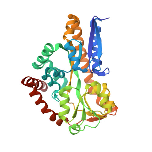
4MMP

Structure of Sialic Acid Binding Protein from Pasturella Multocida


Experimental Data Snapshot

  • Method: X-RAY DIFFRACTION
  • Resolution: 1.57 Å
  • R-Value Free: 0.200 
  • R-Value Work: 0.162 
  • R-Value Observed: 0.164 

Starting Model: experimental
View more details

wwPDB Validation   3D Report Full Report


Ligand Structure Quality Assessment 


This is version 1.3 of the entry. See complete history


Literature

Bacterial periplasmic sialic acid-binding proteins exhibit a conserved binding site.

Gangi Setty, T.Cho, C.Govindappa, S.Apicella, M.A.Ramaswamy, S.

(2014) Acta Crystallogr D Biol Crystallogr 70: 1801-1811

  • DOI: https://doi.org/10.1107/S139900471400830X
  • Primary Citation of Related Structures:  
    4MAG, 4MMP, 4MNP

  • PubMed Abstract: 

    Sialic acids are a family of related nine-carbon sugar acids that play important roles in both eukaryotes and prokaryotes. These sialic acids are incorporated/decorated onto lipooligosaccharides as terminal sugars in multiple bacteria to evade the host immune system. Many pathogenic bacteria scavenge sialic acids from their host and use them for molecular mimicry. The first step of this process is the transport of sialic acid to the cytoplasm, which often takes place using a tripartite ATP-independent transport system consisting of a periplasmic binding protein and a membrane transporter. In this paper, the structural characterization of periplasmic binding proteins from the pathogenic bacteria Fusobacterium nucleatum, Pasteurella multocida and Vibrio cholerae and their thermodynamic characterization are reported. The binding affinities of several mutations in the Neu5Ac binding site of the Haemophilus influenzae protein are also reported. The structure and the thermodynamics of the binding of sugars suggest that all of these proteins have a very well conserved binding pocket and similar binding affinities. A significant conformational change occurs when these proteins bind the sugar. While the C1 carboxylate has been identified as the primary binding site, a second conserved hydrogen-bonding network is involved in the initiation and stabilization of the conformational states.


  • Organizational Affiliation

    Institute for Stem Cell Biology and Regenerative Medicine, NCBS Campus, GKVK Post, Bangalore, Karnataka 560 065, India.


Macromolecules
Find similar proteins by:  (by identity cutoff)  |  3D Structure
Entity ID: 1
MoleculeChains Sequence LengthOrganismDetailsImage
Sialic Acid Binding Protein308Pasteurella multocida subsp. gallicida P1059Mutation(s): 0 
Gene Names: SiaPP1059_01877
Entity Groups  
Sequence Clusters30% Identity50% Identity70% Identity90% Identity95% Identity100% Identity
Sequence Annotations
Expand
  • Reference Sequence
Small Molecules
Ligands 1 Unique
IDChains Name / Formula / InChI Key2D Diagram3D Interactions
SLB
Query on SLB

Download Ideal Coordinates CCD File 
B [auth A]N-acetyl-beta-neuraminic acid
C11 H19 N O9
SQVRNKJHWKZAKO-PFQGKNLYSA-N
Experimental Data & Validation

Experimental Data

  • Method: X-RAY DIFFRACTION
  • Resolution: 1.57 Å
  • R-Value Free: 0.200 
  • R-Value Work: 0.162 
  • R-Value Observed: 0.164 
  • Space Group: P 21 21 21
Unit Cell:
Length ( Å )Angle ( ˚ )
a = 42.643α = 90
b = 77.758β = 90
c = 85.519γ = 90
Software Package:
Software NamePurpose
SCALAdata scaling
AMoREphasing
REFMACrefinement
PDB_EXTRACTdata extraction
HKL-3000data collection
XDSdata reduction

Structure Validation

View Full Validation Report


Ligand Structure Quality Assessment 


Entry History 

Deposition Data

Revision History  (Full details and data files)

  • Version 1.0: 2014-07-09
    Type: Initial release
  • Version 1.1: 2014-09-24
    Changes: Database references
  • Version 1.2: 2020-07-29
    Type: Remediation
    Reason: Carbohydrate remediation
    Changes: Data collection, Database references, Derived calculations, Structure summary
  • Version 1.3: 2023-09-20
    Changes: Data collection, Database references, Refinement description, Structure summary