4MJV | pdb_00004mjv

Influenza Neuraminidase in complex with a novel antiviral compound


Experimental Data Snapshot

  • Method: X-RAY DIFFRACTION
  • Resolution: 2.65 Å
  • R-Value Free: 
    0.285 (Depositor), 0.288 (DCC) 
  • R-Value Work: 
    0.200 (Depositor), 0.205 (DCC) 
  • R-Value Observed: 
    0.208 (Depositor) 

wwPDB Validation 3D Report Full Report

Validation slider image for 4MJV

Ligand Structure Quality Assessment 


This is version 1.3 of the entry. See complete history

Literature

Serendipitous discovery of a potent influenza virus a neuraminidase inhibitor.

Mohan, S.Kerry, P.S.Bance, N.Niikura, M.Pinto, B.M.

(2014) Angew Chem Int Ed Engl 53: 1076-1080

  • DOI: https://doi.org/10.1002/anie.201308142
  • Primary Citation Related Structures: 
    4MJU, 4MJV

  • PubMed Abstract: 

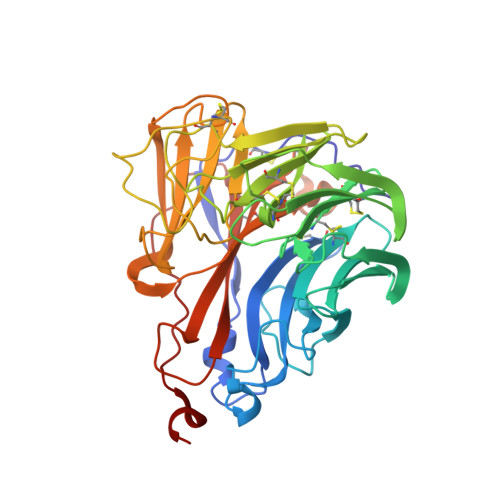
    We have previously reported a potent neuraminidase inhibitor that comprises a carbocyclic analogue of zanamivir in which the hydrophilic glycerol side chain is replaced by the hydrophobic 3-pentyloxy group of oseltamivir. This hybrid inhibitor showed excellent inhibitory properties in the neuraminidase inhibition assay (Ki =0.46 nM; Ki (zanamivir) =0.16 nM) and in the viral replication inhibition assay in cell culture at 10(-8)  M. As part of this lead optimization, we now report a novel spirolactam that shows comparable inhibitory activity in the cell culture assay to that of our lead compound at 10(-7)  M. The compound was discovered serendipitously during the attempted synthesis of the isothiourea derivative of the original candidate. The X-ray crystal structure of the spirolactam in complex with the N8 subtype neuraminidase offers insight into the mode of inhibition.


  • Organizational Affiliation
    • Department of Chemistry, Simon Fraser University, 8888 University Drive, Burnaby, BC, V5A1S6 (Canada) http://www.sfu.ca/chemistry/groups/pinto/

Macromolecule Content 

  • Total Structure Weight: 43.59 kDa 
  • Atom Count: 3,212 
  • Modeled Residue Count: 390 
  • Deposited Residue Count: 390 
  • Unique protein chains: 1

Macromolecules

Find similar proteins by:|  3D Structure
Entity ID: 1
MoleculeChains  Sequence LengthOrganismDetailsImage
Neuraminidase390Influenza A virus (A/duck/Ukraine/1/1963(H3N8))Mutation(s): 0 
EC: 3.2.1.18
UniProt
Find proteins for Q07599 (Influenza A virus (strain A/Duck/Ukraine/1/1963 H3N8))
Explore Q07599 
Go to UniProtKB:  Q07599
Entity Groups
Sequence Clusters30% Identity50% Identity70% Identity90% Identity95% Identity100% Identity
UniProt GroupQ07599
Sequence Annotations
Expand
Reference Sequence

Small Molecules

Ligands 2 Unique
IDChains Name / Formula / InChI Key2D Diagram3D Interactions
27V

Query on 27V


Download:Ideal Coordinates CCD File
B [auth A](2E,5S,9R,10S)-10-(acetylamino)-2-imino-4-oxo-9-(pentan-3-yloxy)-1-thia-3-azaspiro[4.5]dec-6-ene-7-carboxylic acid
C16 H23 N3 O5 S
YPLGHUGDNRYXJS-WQGACYEGSA-N
CA

Query on CA


Download:Ideal Coordinates CCD File
C [auth A]CALCIUM ION
Ca
BHPQYMZQTOCNFJ-UHFFFAOYSA-N

Experimental Data & Validation

Experimental Data

  • Method: X-RAY DIFFRACTION
  • Resolution: 2.65 Å
  • R-Value Free:  0.285 (Depositor), 0.288 (DCC) 
  • R-Value Work:  0.200 (Depositor), 0.205 (DCC) 
  • R-Value Observed: 0.208 (Depositor) 
Space Group: I 4
Unit Cell:
Length ( Å )Angle ( ˚ )
a = 90.744α = 90
b = 90.744β = 90
c = 112.002γ = 90
Software Package:
Software NamePurpose
DENZOdata reduction
SCALEPACKdata scaling
PHASERphasing
PHENIXrefinement
PDB_EXTRACTdata extraction

Structure Validation

View Full Validation Report


Ligand Structure Quality Assessment 


Entry History 

Deposition Data

  • Released Date: 2014-01-15 
  • Deposition Author(s): Kerry, P.S.

Revision History  (Full details and data files)

  • Version 1.0: 2014-01-15
    Type: Initial release
  • Version 1.1: 2014-02-19
    Changes: Database references
  • Version 1.2: 2017-11-15
    Changes: Refinement description
  • Version 1.3: 2024-11-20
    Changes: Data collection, Database references, Derived calculations, Structure summary