4MD9

Crystal Structure of symmetric CK2 holoenzyme with mutated alpha subunit (F121E truncated at aa 336)


Experimental Data Snapshot

  • Method: X-RAY DIFFRACTION
  • Resolution: 3.50 Å
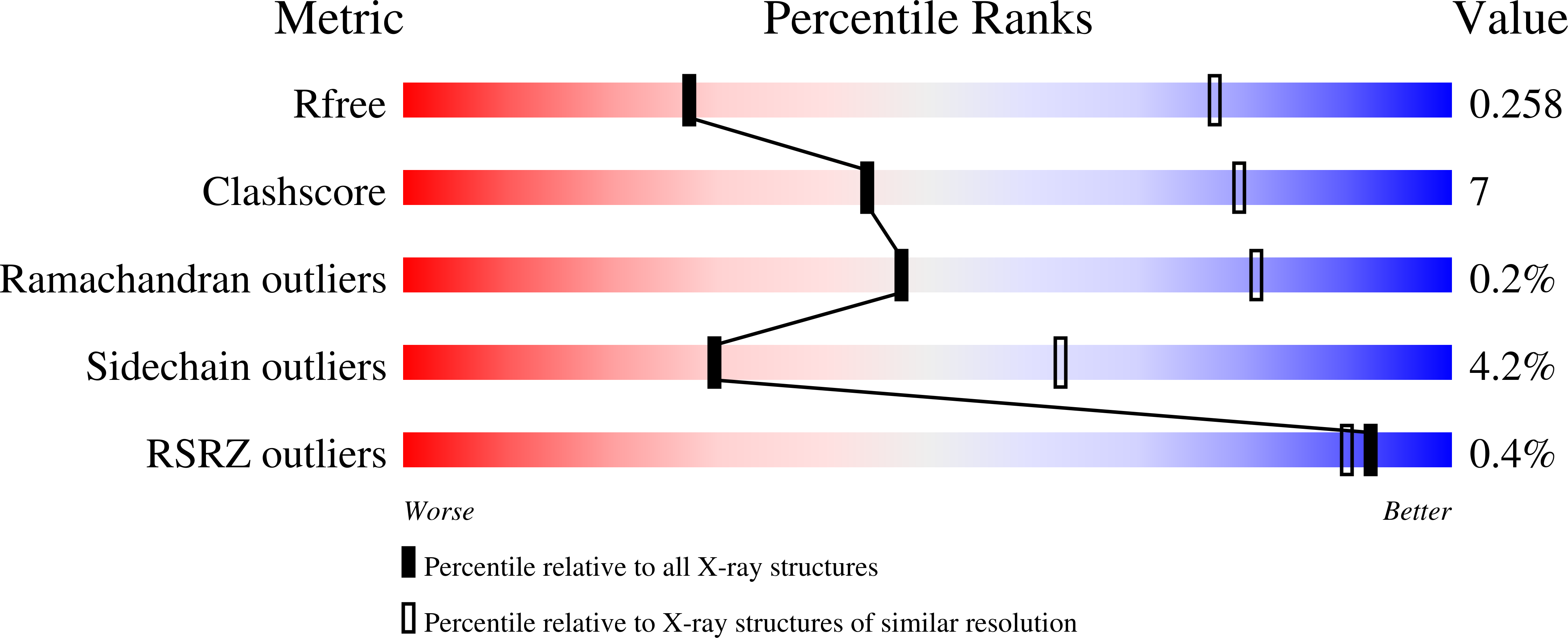
  • R-Value Free: 0.259 
  • R-Value Work: 0.229 
  • R-Value Observed: 0.230 

Starting Models: experimental
View more details

wwPDB Validation   3D Report Full Report


This is version 1.2 of the entry. See complete history


Literature

Active Form of the Protein Kinase CK2 alpha 2 beta 2 Holoenzyme Is a Strong Complex with Symmetric Architecture.

Lolli, G.Ranchio, A.Battistutta, R.

(2014) ACS Chem Biol 9: 366-371

  • DOI: https://doi.org/10.1021/cb400771y
  • Primary Citation of Related Structures:  
    4MD7, 4MD8, 4MD9

  • PubMed Abstract: 

    CK2 is a protein kinase essential for cell viability whose activity is altered in several cancers. Its mechanisms of regulation differ from those common to other eukaryotic protein kinases and are not entirely established yet. Here we present crystal structures of the monomeric form of the α2β2 holoenzyme that allow refining a formerly proposed structural model for activity regulation by oligomerization. Previous crystal structures of the CK2 holoenzyme show an asymmetric arrangement of the two α catalytic subunits around the obligate β2 regulatory subunits. Asymmetric α2β2 tetramers are organized in trimeric rings that correspond to inactive forms of the enzyme. The new crystal structures presented here reveal the symmetric architecture of the isolated active tetramers. The dimension and the nature of the α/β interfaces configure the holoenzyme as a strong complex that does not spontaneously dissociate in solution, in accordance with the low dissociation constant (∼4 nM).


  • Organizational Affiliation

    Department of Chemical Sciences, University of Padua , via Marzolo 1, 35131 Padova, Italy.


Macromolecules
Find similar proteins by:  (by identity cutoff)  |  3D Structure
Entity ID: 1
MoleculeChains Sequence LengthOrganismDetailsImage
Casein kinase II subunit beta
A, B, C, D, I
A, B, C, D, I, J, N, O
215Homo sapiensMutation(s): 0 
Gene Names: CSNK2BCK2NG5A
UniProt & NIH Common Fund Data Resources
Find proteins for P67870 (Homo sapiens)
Explore P67870 
Go to UniProtKB:  P67870
PHAROS:  P67870
GTEx:  ENSG00000204435 
Entity Groups  
Sequence Clusters30% Identity50% Identity70% Identity90% Identity95% Identity100% Identity
UniProt GroupP67870
Sequence Annotations
Expand
  • Reference Sequence
Find similar proteins by:  (by identity cutoff)  |  3D Structure
Entity ID: 2
MoleculeChains Sequence LengthOrganismDetailsImage
Casein kinase II subunit alpha
E, F, G, H, K
E, F, G, H, K, L, M, P
336Homo sapiensMutation(s): 1 
Gene Names: CSNK2A1CK2A1
EC: 2.7.11.1
UniProt & NIH Common Fund Data Resources
Find proteins for P68400 (Homo sapiens)
Explore P68400 
Go to UniProtKB:  P68400
PHAROS:  P68400
GTEx:  ENSG00000101266 
Entity Groups  
Sequence Clusters30% Identity50% Identity70% Identity90% Identity95% Identity100% Identity
UniProt GroupP68400
Sequence Annotations
Expand
  • Reference Sequence
Experimental Data & Validation

Experimental Data

  • Method: X-RAY DIFFRACTION
  • Resolution: 3.50 Å
  • R-Value Free: 0.259 
  • R-Value Work: 0.229 
  • R-Value Observed: 0.230 
  • Space Group: C 1 2 1
Unit Cell:
Length ( Å )Angle ( ˚ )
a = 283.924α = 90
b = 115.707β = 119.22
c = 208.223γ = 90
Software Package:
Software NamePurpose
SCALAdata scaling
PHASERphasing
REFMACrefinement
PDB_EXTRACTdata extraction
XSCALEdata scaling

Structure Validation

View Full Validation Report


Entry History 

Deposition Data

Revision History  (Full details and data files)

  • Version 1.0: 2013-11-20
    Type: Initial release
  • Version 1.1: 2014-03-05
    Changes: Database references
  • Version 1.2: 2023-09-20
    Changes: Data collection, Database references, Derived calculations, Refinement description