4MCY | pdb_00004mcy

Immune Receptor


Experimental Data Snapshot

  • Method: X-RAY DIFFRACTION
  • Resolution: 2.30 Å
  • R-Value Free: 
    0.225 (Depositor), 0.221 (DCC) 
  • R-Value Work: 
    0.171 (Depositor), 0.174 (DCC) 
  • R-Value Observed: 
    0.173 (Depositor) 

wwPDB Validation 3D Report Full Report

Validation slider image for 4MCY

Ligand Structure Quality Assessment 


This is version 2.1 of the entry. See complete history. 

Literature

A molecular basis for the association of the HLA-DRB1 locus, citrullination, and rheumatoid arthritis.

Scally, S.W., Petersen, J., Law, S.C., Dudek, N.L., Nel, H.J., Loh, K.L., Wijeyewickrema, L.C., Eckle, S.B., van Heemst, J., Pike, R.N., McCluskey, J., Toes, R.E., La Gruta, N.L., Purcell, A.W., Reid, H.H., Thomas, R., Rossjohn, J.

(2013) J Exp Med 210: 2569-2582

  • DOI: https://doi.org/10.1084/jem.20131241
  • Primary Citation Related Structures: 
    4MCY, 4MCZ, 4MD0, 4MD4, 4MD5, 4MDI, 4MDJ

  • PubMed Abstract: 

    Rheumatoid arthritis (RA) is strongly associated with the human leukocyte antigen (HLA)-DRB1 locus that possesses the shared susceptibility epitope (SE) and the citrullination of self-antigens. We show how citrullinated aggrecan and vimentin epitopes bind to HLA-DRB1*04:01/04. Citrulline was accommodated within the electropositive P4 pocket of HLA-DRB1*04:01/04, whereas the electronegative P4 pocket of the RA-resistant HLA-DRB1*04:02 allomorph interacted with arginine or citrulline-containing epitopes. Peptide elution studies revealed P4 arginine-containing peptides from HLA-DRB1*04:02, but not from HLA-DRB1*04:01/04. Citrullination altered protease susceptibility of vimentin, thereby generating self-epitopes that are presented to T cells in HLA-DRB1*04:01(+) individuals. Using HLA-II tetramers, we observed citrullinated vimentin- and aggrecan-specific CD4(+) T cells in the peripheral blood of HLA-DRB1*04:01(+) RA-affected and healthy individuals. In RA patients, autoreactive T cell numbers correlated with disease activity and were deficient in regulatory T cells relative to healthy individuals. These findings reshape our understanding of the association between citrullination, the HLA-DRB1 locus, and T cell autoreactivity in RA.


  • Organizational Affiliation: 
    • Department of Biochemistry and Molecular Biology, School of Biomedical Sciences, Monash University, Clayton, Victoria 3800, Australia.

Macromolecule Content 

  • Total Structure Weight: 47.32 kDa 
  • Atom Count: 3,500 
  • Modeled Residue Count: 381 
  • Deposited Residue Count: 402 
  • Unique protein chains: 3

Macromolecules

Find similar proteins by:|  3D Structure
Entity ID: 1
MoleculeChains  Sequence LengthOrganismDetailsImage
HLA class II histocompatibility antigen, DR alpha chain189Homo sapiensMutation(s): 0 
Gene Names: HLA-DRA, HLA-DRA1
UniProt & NIH Common Fund Data Resources
Find proteins for P01903 (Homo sapiens)
Explore P01903 
Go to UniProtKB:  P01903
PHAROS:  P01903
GTEx:  ENSG00000204287 
Entity Groups
Sequence Clusters30% Identity50% Identity70% Identity90% Identity95% Identity100% Identity
UniProt GroupP01903
Glycosylation
Glycosylation Sites: 2Go to GlyGen: P01903-1
Sequence Annotations
Expand
Reference Sequence
Find similar proteins by:|  3D Structure
Entity ID: 2
MoleculeChains  Sequence LengthOrganismDetailsImage
HLA class II histocompatibility antigen, DRB1-4 beta chain200Homo sapiensMutation(s): 0 
Gene Names: HLA-DRB1
UniProt & NIH Common Fund Data Resources
Find proteins for P01911 (Homo sapiens)
Explore P01911 
Go to UniProtKB:  P01911
PHAROS:  P01911
GTEx:  ENSG00000196126 
Entity Groups
Sequence Clusters30% Identity50% Identity70% Identity90% Identity95% Identity100% Identity
UniProt GroupP01911
Glycosylation
Glycosylation Sites: 1Go to GlyGen: P01911-1
Sequence Annotations
Expand
Reference Sequence
Find similar proteins by:  Sequence   |   3D Structure  
Entity ID: 3
MoleculeChains  Sequence LengthOrganismDetailsImage
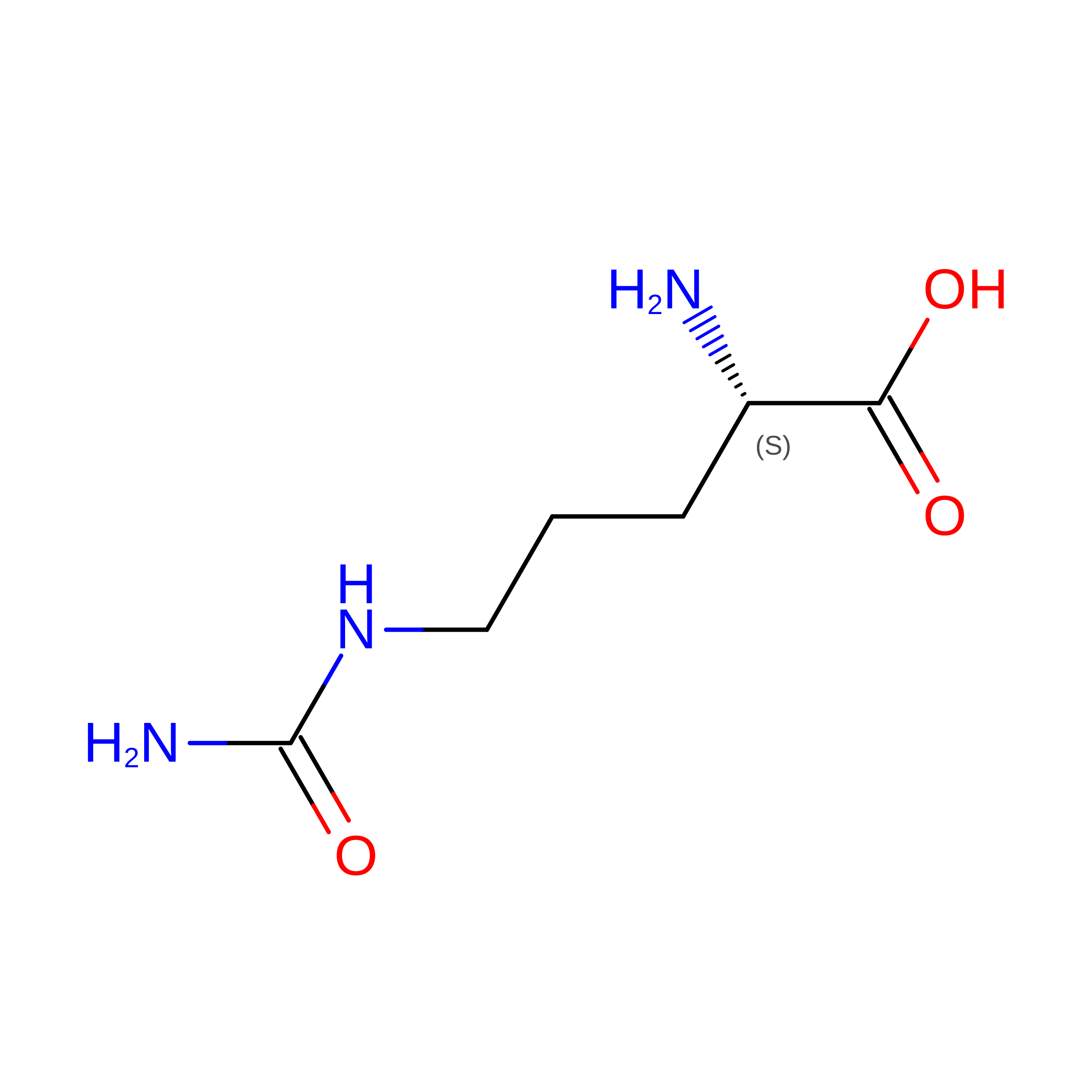
Citrullinated Vimentin13Homo sapiensMutation(s): 0 
UniProt & NIH Common Fund Data Resources
Find proteins for P08670 (Homo sapiens)
Explore P08670 
Go to UniProtKB:  P08670
PHAROS:  P08670
GTEx:  ENSG00000026025 
Entity Groups
Sequence Clusters30% Identity50% Identity70% Identity90% Identity95% Identity100% Identity
UniProt GroupP08670
Sequence Annotations
Expand
Reference Sequence

Experimental Data & Validation

Experimental Data

  • Method: X-RAY DIFFRACTION
  • Resolution: 2.30 Å
  • R-Value Free:  0.225 (Depositor), 0.221 (DCC) 
  • R-Value Work:  0.171 (Depositor), 0.174 (DCC) 
  • R-Value Observed: 0.173 (Depositor) 
Space Group: C 2 2 21
Unit Cell:
Length ( Å )Angle ( ˚ )
a = 67.035α = 90
b = 177.821β = 90
c = 76.728γ = 90
Software Package:
Software NamePurpose
Blu-Icedata collection
PHASERphasing
PHENIXrefinement
MOSFLMdata reduction
SCALAdata scaling

Structure Validation

View Full Validation Report


Ligand Structure Quality Assessment 


Entry History 

Deposition Data

Revision History  (Full details and data files)

  • Version 1.0: 2013-11-27
    Type: Initial release
  • Version 1.1: 2013-12-04
    Changes: Database references
  • Version 1.2: 2020-07-29
    Type: Remediation
    Reason: Carbohydrate remediation
    Changes: Data collection, Database references, Derived calculations, Structure summary
  • Version 2.0: 2023-11-15
    Changes: Atomic model, Data collection, Database references, Derived calculations, Structure summary
  • Version 2.1: 2024-11-20
    Changes: Structure summary