4LZ8 | pdb_00004lz8

Crystal structures of GLuR2 ligand-binding-domain in complex with glutamate and positive allosteric modulators


Experimental Data Snapshot

  • Method: X-RAY DIFFRACTION
  • Resolution: 1.85 Å
  • R-Value Free: 
    0.232 (Depositor), 0.218 (DCC) 
  • R-Value Work: 
    0.196 (Depositor) 
  • R-Value Observed: 
    0.198 (Depositor) 

Starting Model: experimental
View more details

wwPDB Validation 3D Report Full Report

Validation slider image for 4LZ8

Ligand Structure Quality Assessment 


This is version 1.4 of the entry. See complete history

Literature

Discovery and Characterization of a Novel Dihydroisoxazole Class of alpha-Amino-3-hydroxy-5-methyl-4-isoxazolepropionic acid (AMPA) Receptor Potentiators.

Patel, N.C.Schwarz, J.Hou, X.J.Hoover, D.J.Xie, L.Fliri, A.J.Gallaschun, R.J.Lazzaro, J.T.Bryce, D.K.Hoffmann, W.E.Hanks, A.N.McGinnis, D.Marr, E.S.Gazard, J.L.Hajos, M.Scialis, R.J.Hurst, R.S.Shaffer, C.L.Pandit, J.O'Donnell, C.J.

(2013) J Med Chem 56: 9180-9191

  • DOI: https://doi.org/10.1021/jm401274b
  • Primary Citation Related Structures: 
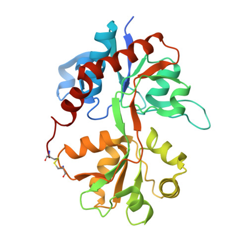
    4LZ5, 4LZ7, 4LZ8

  • PubMed Abstract: 

    Positive allosteric modulators ("potentiators") of α-amino-3-hydroxy-5-methyl-4-isoxazolepropionic acid (AMPA) receptors (AMPAR) enhance excitatory neurotransmission and may improve the cognitive deficits associated with various neurological disorders. The dihydroisoxazole (DHI) series of AMPAR potentiators described herein originated from the identification of 7 by a high-throughput functional activity screen using mouse embryonic stem (mES) cell-derived neuronal precursors. Subsequent structure-based drug design using X-ray crystal structures of the ligand-binding domain of human GluA2 led to the discovery of both PF-04725379 (11), which in tritiated form became a novel ligand for characterizing the binding affinities of subsequent AMPAR potentiators in rat brain homogenate, and PF-04701475 (8a), a prototype used to explore AMPAR-mediated pharmacology in vivo. Lead series optimization provided 16a, a functionally potent compound lacking the potentially bioactivatable aniline within 8a, but retaining desirable in vitro ADME properties.


  • Organizational Affiliation
    • Pfizer Worldwide Research and Development, Groton Laboratories , 445 Eastern Point Road, Groton, Connecticut 06340, United States.

Macromolecule Content 

  • Total Structure Weight: 92.65 kDa 
  • Atom Count: 6,503 
  • Modeled Residue Count: 772 
  • Deposited Residue Count: 825 
  • Unique protein chains: 1

Macromolecules

Find similar proteins by:|  3D Structure
Entity ID: 1
MoleculeChains  Sequence LengthOrganismDetailsImage
Glutamate receptor 2
A, B, C
275Rattus norvegicusMutation(s): 3 
Gene Names: Gria2Glur2
UniProt
Find proteins for P19491 (Rattus norvegicus)
Explore P19491 
Go to UniProtKB:  P19491
Entity Groups
Sequence Clusters30% Identity50% Identity70% Identity90% Identity95% Identity100% Identity
UniProt GroupP19491
Sequence Annotations
Expand
Reference Sequence

Small Molecules

Ligands 3 Unique
IDChains Name / Formula / InChI Key2D Diagram3D Interactions
1YX

Query on 1YX


Download:Ideal Coordinates CCD File
E [auth A],
J [auth B]
N-[(3S)-1-{2-fluoro-4-[(5S)-5-{[(propan-2-ylsulfonyl)amino]methyl}-4,5-dihydro-1,2-oxazol-3-yl]phenyl}pyrrolidin-3-yl]acetamide
C19 H27 F N4 O4 S
XCANXEILGPMNGT-HOTGVXAUSA-N
GLU

Query on GLU


Download:Ideal Coordinates CCD File
D [auth A],
I [auth B],
M [auth C]
GLUTAMIC ACID
C5 H9 N O4
WHUUTDBJXJRKMK-VKHMYHEASA-N
ZN

Query on ZN


Download:Ideal Coordinates CCD File
F [auth B],
G [auth B],
H [auth B],
K [auth C],
L [auth C]
ZINC ION
Zn
PTFCDOFLOPIGGS-UHFFFAOYSA-N

Experimental Data & Validation

Experimental Data

  • Method: X-RAY DIFFRACTION
  • Resolution: 1.85 Å
  • R-Value Free:  0.232 (Depositor), 0.218 (DCC) 
  • R-Value Work:  0.196 (Depositor) 
  • R-Value Observed: 0.198 (Depositor) 
Space Group: P 21 21 2
Unit Cell:
Length ( Å )Angle ( ˚ )
a = 113.676α = 90
b = 164.953β = 90
c = 47.498γ = 90
Software Package:
Software NamePurpose
DENZOdata reduction
SCALEPACKdata scaling
REFMACrefinement
PDB_EXTRACTdata extraction
AMoREphasing

Structure Validation

View Full Validation Report


Ligand Structure Quality Assessment 


Entry History 

Deposition Data

  • Released Date: 2013-12-04 
  • Deposition Author(s): Pandit, J.

Revision History  (Full details and data files)

  • Version 1.0: 2013-12-04
    Type: Initial release
  • Version 1.1: 2013-12-11
    Changes: Database references
  • Version 1.2: 2017-07-26
    Changes: Refinement description, Source and taxonomy
  • Version 1.3: 2023-09-20
    Changes: Data collection, Database references, Derived calculations, Refinement description
  • Version 1.4: 2024-11-27
    Changes: Structure summary