4LXQ | pdb_00004lxq

Crystal structure WlaRD, a sugar 3N-formyl transferase in the presence of dTdp and 5-N-Formyl-THF


Experimental Data Snapshot

  • Method: X-RAY DIFFRACTION
  • Resolution: 1.40 Å
  • R-Value Free: 
    0.178 (Depositor), 0.177 (DCC) 
  • R-Value Work: 
    0.158 (Depositor), 0.157 (DCC) 
  • R-Value Observed: 
    0.159 (Depositor) 

Starting Model: experimental
View more details

wwPDB Validation 3D Report Full Report

Validation slider image for 4LXQ

Ligand Structure Quality Assessment 


This is version 1.2 of the entry. See complete history

Literature

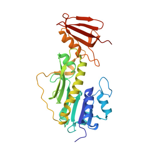
Structure of a sugar N-formyltransferase from Campylobacter jejuni.

Thoden, J.B.Goneau, M.F.Gilbert, M.Holden, H.M.

(2013) Biochemistry 52: 6114-6126

  • DOI: https://doi.org/10.1021/bi4009006
  • Primary Citation Related Structures: 
    4LXQ, 4LXT, 4LXU, 4LXX, 4LXY, 4LY0, 4LY3

  • PubMed Abstract: 

    The O-antigens, which are components of the outer membranes of Gram-negative bacteria, are responsible for the wide species variations seen in nature and are thought to play a role in bacterial virulence. They often contain unusual dideoxysugars such as 3,6-dideoxy-3-formamido-d-glucose (Qui3NFo). Here, we describe a structural and functional investigation of the protein C8J_1081 from Campylobacter jejuni 81116, which is involved in the biosynthesis of Qui3NFo. Specifically, the enzyme, hereafter referred to as WlaRD, catalyzes the N-formylation of dTDP-3,6-dideoxy-3-amino-d-glucose (dTDP-Qui3N) using N(10)-formyltetrahydrofolate as the carbon source. For this investigation, seven X-ray structures of WlaRD, in complexes with various dTDP-linked sugars and cofactors, were determined to resolutions of 1.9 Å or better. One of the models, with bound N(10)-formyltetrahydrofolate and dTDP, represents the first glimpse of an N-formyltransferase with its natural cofactor. Another model contains the reaction products, tetrahydrofolate and dTDP-Qui3NFo. In combination, the structures provide snapshots of the WlaRD active site before and after catalysis. On the basis of these structures, three amino acid residues were targeted for study: Asn 94, His 96, and Asp 132. Mutations of any of these residues resulted in a complete loss of enzymatic activity. Given the position of His 96 in the active site, it can be postulated that it functions as the active site base to remove a proton from the sugar amino group as it attacks the carbonyl carbon of the N-10 formyl group of the cofactor. Enzyme assays demonstrate that WlaRD is also capable of utilizing dTDP-3,6-dideoxy-3-amino-d-galactose (dTDP-Fuc3N) as a substrate, albeit at a much reduced catalytic efficiency.


  • Organizational Affiliation
    • Department of Biochemistry, University of Wisconsin , Madison, Wisconsin 53706, United States.

Macromolecule Content 

  • Total Structure Weight: 67.39 kDa 
  • Atom Count: 5,890 
  • Modeled Residue Count: 547 
  • Deposited Residue Count: 548 
  • Unique protein chains: 1

Macromolecules

Find similar proteins by:|  3D Structure
Entity ID: 1
MoleculeChains  Sequence LengthOrganismDetailsImage
WlaRD, a sugar 3N-formyl transferase
A, B
274Campylobacter jejuni subsp. jejuni 81116Mutation(s): 0 
Gene Names: C8J_1081

Small Molecules

Ligands 5 Unique
IDChains Name / Formula / InChI Key2D Diagram3D Interactions
FON

Query on FON


Download:Ideal Coordinates CCD File
C [auth A],
N [auth B]
N-{[4-({[(6R)-2-amino-5-formyl-4-oxo-1,4,5,6,7,8-hexahydropteridin-6-yl]methyl}amino)phenyl]carbonyl}-L-glutamic acid
C20 H23 N7 O7
VVIAGPKUTFNRDU-OLZOCXBDSA-N
TYD

Query on TYD


Download:Ideal Coordinates CCD File
D [auth A],
O [auth B]
THYMIDINE-5'-DIPHOSPHATE
C10 H16 N2 O11 P2
UJLXYODCHAELLY-XLPZGREQSA-N
MPO

Query on MPO


Download:Ideal Coordinates CCD File
E [auth A],
H [auth A],
P [auth B],
Q [auth B]
3[N-MORPHOLINO]PROPANE SULFONIC ACID
C7 H15 N O4 S
DVLFYONBTKHTER-UHFFFAOYSA-N
EDO

Query on EDO


Download:Ideal Coordinates CCD File
AA [auth B]
BA [auth B]
F [auth A]
G [auth A]
I [auth A]
AA [auth B],
BA [auth B],
F [auth A],
G [auth A],
I [auth A],
R [auth B],
S [auth B],
T [auth B],
U [auth B],
Y [auth B],
Z [auth B]
1,2-ETHANEDIOL
C2 H6 O2
LYCAIKOWRPUZTN-UHFFFAOYSA-N
CL

Query on CL


Download:Ideal Coordinates CCD File
J [auth A]
K [auth A]
L [auth A]
M [auth A]
V [auth B]
J [auth A],
K [auth A],
L [auth A],
M [auth A],
V [auth B],
W [auth B],
X [auth B]
CHLORIDE ION
Cl
VEXZGXHMUGYJMC-UHFFFAOYSA-M

Experimental Data & Validation

Experimental Data

  • Method: X-RAY DIFFRACTION
  • Resolution: 1.40 Å
  • R-Value Free:  0.178 (Depositor), 0.177 (DCC) 
  • R-Value Work:  0.158 (Depositor), 0.157 (DCC) 
  • R-Value Observed: 0.159 (Depositor) 
Space Group: C 1 2 1
Unit Cell:
Length ( Å )Angle ( ˚ )
a = 97.861α = 90
b = 63.259β = 110.04
c = 136.212γ = 90
Software Package:
Software NamePurpose
HKL-3000data collection
PHASERphasing
REFMACrefinement
HKL-3000data reduction
HKL-3000data scaling

Structure Validation

View Full Validation Report


Ligand Structure Quality Assessment 


Entry History 

Deposition Data

Revision History  (Full details and data files)

  • Version 1.0: 2013-08-14
    Type: Initial release
  • Version 1.1: 2014-02-05
    Changes: Database references
  • Version 1.2: 2023-09-20
    Changes: Data collection, Database references, Derived calculations, Refinement description