4LWP | pdb_00004lwp

Crystal structure of PRMT6-SAH


Experimental Data Snapshot

  • Method: X-RAY DIFFRACTION
  • Resolution: 2.35 Å
  • R-Value Free: 
    0.249 (Depositor), 0.248 (DCC) 
  • R-Value Work: 
    0.204 (Depositor), 0.206 (DCC) 
  • R-Value Observed: 
    0.207 (Depositor) 

wwPDB Validation 3D Report Full Report

Validation slider image for 4LWP

Ligand Structure Quality Assessment 


This is version 1.1 of the entry. See complete history

Literature

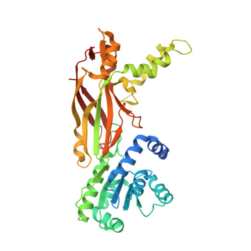
Crystal Structure of Arginine Methyltransferase 6 from Trypanosoma brucei

Wang, C.Zhu, Y.Chen, J.Li, X.Peng, J.Chen, J.Zou, Y.Zhang, Z.Jin, H.Yang, P.Wu, J.Niu, L.Gong, Q.Teng, M.Shi, Y.

(2014) PLoS One 9: e87267-e87267

  • DOI: https://doi.org/10.1371/journal.pone.0087267
  • Primary Citation Related Structures: 
    4LWO, 4LWP

  • PubMed Abstract: 

    Arginine methylation plays vital roles in the cellular functions of the protozoan Trypanosoma brucei. The T. brucei arginine methyltransferase 6 (TbPRMT6) is a type I arginine methyltransferase homologous to human PRMT6. In this study, we report the crystal structures of apo-TbPRMT6 and its complex with the reaction product S-adenosyl-homocysteine (SAH). The structure of apo-TbPRMT6 displays several features that are different from those of type I PRMTs that were structurally characterized previously, including four stretches of insertion, the absence of strand β15, and a distinct dimerization arm. The comparison of the apo-TbPRMT6 and SAH-TbPRMT6 structures revealed the fine rearrangements in the active site upon SAH binding. The isothermal titration calorimetry results demonstrated that SAH binding greatly increases the affinity of TbPRMT6 to a substrate peptide derived from bovine histone H4. The western blotting and mass spectrometry results revealed that TbPRMT6 methylates bovine histone H4 tail at arginine 3 but cannot methylate several T. brucei histone tails. In summary, our results highlight the structural differences between TbPRMT6 and other type I PRMTs and reveal that the active site rearrangement upon SAH binding is important for the substrate binding of TbPRMT6.


  • Organizational Affiliation
    • Hefei National Laboratory for Physical Sciences at Microscale and School of Life Sciences, University of Science and Technology of China, Anhui, China.

Macromolecule Content 

  • Total Structure Weight: 84.58 kDa 
  • Atom Count: 5,312 
  • Modeled Residue Count: 683 
  • Deposited Residue Count: 736 
  • Unique protein chains: 1

Macromolecules

Find similar proteins by:|  3D Structure
Entity ID: 1
MoleculeChains  Sequence LengthOrganismDetailsImage
Arginine N-methyltransferase, putativeA [auth B],
B [auth A]
368Trypanosoma brucei brucei TREU927Mutation(s): 0 
Gene Names: Tb927.5.3960
UniProt
Find proteins for Q57U70 (Trypanosoma brucei brucei (strain 927/4 GUTat10.1))
Explore Q57U70 
Go to UniProtKB:  Q57U70
Entity Groups
Sequence Clusters30% Identity50% Identity70% Identity90% Identity95% Identity100% Identity
UniProt GroupQ57U70
Sequence Annotations
Expand
Reference Sequence

Small Molecules

Ligands 3 Unique
IDChains Name / Formula / InChI Key2D Diagram3D Interactions
SAH

Query on SAH


Download:Ideal Coordinates CCD File
I [auth B],
J [auth A]
S-ADENOSYL-L-HOMOCYSTEINE
C14 H20 N6 O5 S
ZJUKTBDSGOFHSH-WFMPWKQPSA-N
PGE

Query on PGE


Download:Ideal Coordinates CCD File
N [auth A]TRIETHYLENE GLYCOL
C6 H14 O4
ZIBGPFATKBEMQZ-UHFFFAOYSA-N
IOD

Query on IOD


Download:Ideal Coordinates CCD File
C [auth B]
D [auth B]
E [auth B]
F [auth B]
G [auth B]
C [auth B],
D [auth B],
E [auth B],
F [auth B],
G [auth B],
H [auth B],
K [auth A],
L [auth A],
M [auth A]
IODIDE ION
I
XMBWDFGMSWQBCA-UHFFFAOYSA-M

Experimental Data & Validation

Experimental Data

  • Method: X-RAY DIFFRACTION
  • Resolution: 2.35 Å
  • R-Value Free:  0.249 (Depositor), 0.248 (DCC) 
  • R-Value Work:  0.204 (Depositor), 0.206 (DCC) 
  • R-Value Observed: 0.207 (Depositor) 
Space Group: C 2 2 21
Unit Cell:
Length ( Å )Angle ( ˚ )
a = 101.428α = 90
b = 108.465β = 90
c = 137.865γ = 90
Software Package:
Software NamePurpose
HKL-2000data collection
PHENIXmodel building
PHENIXrefinement
HKL-2000data reduction
SCALAdata scaling
PHENIXphasing

Structure Validation

View Full Validation Report


Ligand Structure Quality Assessment 


Entry History 

Deposition Data

Revision History  (Full details and data files)

  • Version 1.0: 2014-02-19
    Type: Initial release
  • Version 1.1: 2024-03-20
    Changes: Data collection, Database references, Derived calculations