4LV7 | pdb_00004lv7

Crystal structure of inositol 1,3,4,5,6-pentakisphosphate 2-kinase E82C/S142C


Experimental Data Snapshot

  • Method: X-RAY DIFFRACTION
  • Resolution: 2.60 Å
  • R-Value Free: 
    0.294 (Depositor), 0.287 (DCC) 
  • R-Value Work: 
    0.224 (Depositor), 0.216 (DCC) 
  • R-Value Observed: 
    0.229 (Depositor) 

Starting Model: experimental
View more details

wwPDB Validation 3D Report Full Report

Validation slider image for 4LV7

Ligand Structure Quality Assessment 


This is version 1.5 of the entry. See complete history

Literature

Conformational stability of inositol 1,3,4,5,6-pentakisphosphate 2-kinase (IPK1) dictates its substrate selectivity.

Gosein, V.Miller, G.J.

(2013) J Biological Chem 288: 36788-36795

  • DOI: https://doi.org/10.1074/jbc.M113.512731
  • Primary Citation Related Structures: 
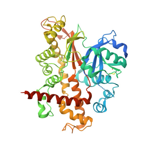
    4LV7

  • PubMed Abstract: 

    Inositol 1,3,4,5,6-pentakisphosphate 2-kinase (IPK1) converts inositol 1,3,4,5,6-pentakisphosphate(IP5) to inositol hexakisphosphate (IP6). IPK1 shares structural similarity with protein kinases and is suspected to employ a similar mechanism of activation. Previous studies revealed roles for the 1- and 3-phosphates of IP5 in IPK1 activation and revealed that the N-lobe of IPK1 is unstable in the absence of inositol phosphate (IP). Here, we demonstrate the link between IPK1 substrate specificity and the stability of its N-lobe. Limited proteolysis of IPK1 revealed that N-lobe stability is dependent on the presence of the 1-phosphate of the substrate, whereas overall stability of IPK1 was increased in ternary complexes with nucleotide and IPs possessing 1- and 3-phosphates that engage the N-lobe of IPK1. Thus, the 1- and 3-phosphates possess dual roles in both IPK1 activation and IPK1 stability. To test whether kinase stability directly contributed to substrate selectivity of the kinase, we engineered IPK1 mutants with disulfide bonds that artificially stabilized the N-lobe in an IP-independent manner thereby mimicking its substrate-bound state in the absence of IP. IPK1 E82C/S142C exhibited a DTT-sensitive 5-fold increase in kcat for 3,4,5,6-inositol tetrakisphosphate (3,4,5,6-IP4) as compared with wild-type IPK1. The crystal structure of the IPK1 E82C/S142C mutant confirmed the presence of the disulfide bond and revealed a small shift in the N-lobe. Finally, we determined that IPK1 E82C/S142C is substantially more stable than wild-type IPK1 under nonreducing conditions, revealing that increased stability of IPK1 E82C/S142C correlates with changes in substrate specificity by allowing IPs lacking the stabilizing 1-phosphate to be used. Taken together, our results show that IPK1 substrate selection is linked to the ability of each potential substrate to stabilize IPK1.


  • Organizational Affiliation
    • From the Department of Pharmacology and Therapeutics, McGill University, Montréal, Quebec H3G 1Y6, Canada.

Macromolecule Content 

  • Total Structure Weight: 113 kDa 
  • Atom Count: 6,380 
  • Modeled Residue Count: 784 
  • Deposited Residue Count: 986 
  • Unique protein chains: 1

Macromolecules

Find similar proteins by:|  3D Structure
Entity ID: 1
MoleculeChains  Sequence LengthOrganismDetailsImage
Inositol-pentakisphosphate 2-kinase
A, B
493Arabidopsis thalianaMutation(s): 2 
Gene Names: At5g42810IPK1MJB21.19
EC: 2.7.1.158
UniProt
Find proteins for Q93YN9 (Arabidopsis thaliana)
Explore Q93YN9 
Go to UniProtKB:  Q93YN9
Entity Groups
Sequence Clusters30% Identity50% Identity70% Identity90% Identity95% Identity100% Identity
UniProt GroupQ93YN9
Sequence Annotations
Expand
Reference Sequence

Small Molecules

Ligands 4 Unique
IDChains Name / Formula / InChI Key2D Diagram3D Interactions
IHP

Query on IHP


Download:Ideal Coordinates CCD File
C [auth A],
G [auth B]
INOSITOL HEXAKISPHOSPHATE
C6 H18 O24 P6
IMQLKJBTEOYOSI-GPIVLXJGSA-N
ADP

Query on ADP


Download:Ideal Coordinates CCD File
D [auth A],
H [auth B]
ADENOSINE-5'-DIPHOSPHATE
C10 H15 N5 O10 P2
XTWYTFMLZFPYCI-KQYNXXCUSA-N
ZN

Query on ZN


Download:Ideal Coordinates CCD File
F [auth A],
J [auth B]
ZINC ION
Zn
PTFCDOFLOPIGGS-UHFFFAOYSA-N
MG

Query on MG


Download:Ideal Coordinates CCD File
E [auth A],
I [auth B]
MAGNESIUM ION
Mg
JLVVSXFLKOJNIY-UHFFFAOYSA-N

Experimental Data & Validation

Experimental Data

  • Method: X-RAY DIFFRACTION
  • Resolution: 2.60 Å
  • R-Value Free:  0.294 (Depositor), 0.287 (DCC) 
  • R-Value Work:  0.224 (Depositor), 0.216 (DCC) 
  • R-Value Observed: 0.229 (Depositor) 
Space Group: P 1
Unit Cell:
Length ( Å )Angle ( ˚ )
a = 59.763α = 83.01
b = 59.353β = 89.92
c = 82.232γ = 63.41
Software Package:
Software NamePurpose
DENZOdata reduction
SCALEPACKdata scaling
PHENIXrefinement
PDB_EXTRACTdata extraction
StructureStudiodata collection
HKL-2000data reduction
HKL-2000data scaling
SOLVEphasing

Structure Validation

View Full Validation Report


Ligand Structure Quality Assessment 


Entry History 

Deposition Data

Revision History  (Full details and data files)

  • Version 1.0: 2013-11-06
    Type: Initial release
  • Version 1.1: 2017-11-15
    Changes: Refinement description
  • Version 1.2: 2018-04-18
    Changes: Data collection, Database references
  • Version 1.3: 2020-10-14
    Changes: Database references, Derived calculations, Structure summary
  • Version 1.4: 2023-09-20
    Changes: Data collection, Database references, Refinement description
  • Version 1.5: 2024-11-20
    Changes: Structure summary