4LSX | pdb_00004lsx

Plant steroid receptor ectodomain bound to brassinolide and SERK1 co-receptor ectodomain


Experimental Data Snapshot

  • Method: X-RAY DIFFRACTION
  • Resolution: 3.30 Å
  • R-Value Free: 
    0.285 (Depositor), 0.281 (DCC) 
  • R-Value Work: 
    0.246 (Depositor), 0.241 (DCC) 
  • R-Value Observed: 
    0.248 (Depositor) 

Starting Models: experimental
View more details

wwPDB Validation 3D Report Full Report

Validation slider image for 4LSX

Ligand Structure Quality Assessment 


This is version 2.2 of the entry. See complete history

Literature

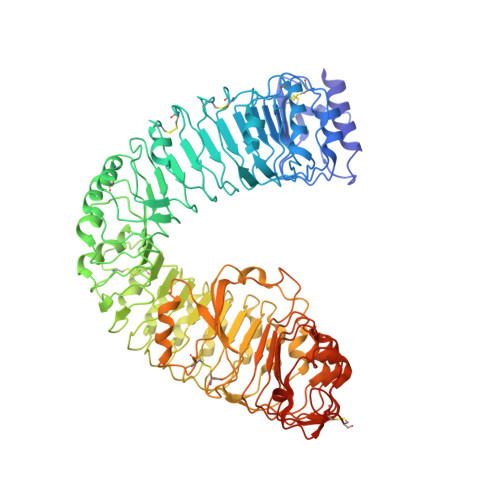
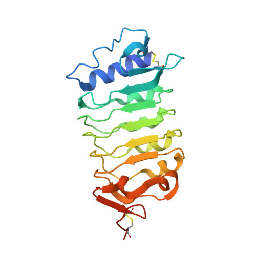
Molecular mechanism for plant steroid receptor activation by somatic embryogenesis co-receptor kinases.

Santiago, J.Henzler, C.Hothorn, M.

(2013) Science 341: 889-892

  • DOI: https://doi.org/10.1126/science.1242468
  • Primary Citation Related Structures: 
    4LSA, 4LSC, 4LSX

  • PubMed Abstract: 

    Brassinosteroids, which control plant growth and development, are sensed by the leucine-rich repeat (LRR) domain of the membrane receptor kinase BRASSINOSTEROID INSENSITIVE 1 (BRI1), but it is unknown how steroid binding at the cell surface activates the cytoplasmic kinase domain of the receptor. A family of somatic embryogenesis receptor kinases (SERKs) has been genetically implicated in mediating early brassinosteroid signaling events. We found a direct and steroid-dependent interaction between the BRI1 and SERK1 LRR domains by analysis of their complex crystal structure at 3.3 angstrom resolution. We show that the SERK1 LRR domain is involved in steroid sensing and, through receptor-co-receptor heteromerization, in the activation of the BRI1 signaling pathway. Our work reveals how known missense mutations in BRI1 and in SERKs modulate brassinosteroid signaling and the targeting mechanism of BRI1 receptor antagonists.


  • Organizational Affiliation
    • Structural Plant Biology Lab, Friedrich Miescher Laboratory of the Max Planck Society, Spemannstraße 39, Tübingen 72076, Germany.

Macromolecule Content 

  • Total Structure Weight: 218.28 kDa 
  • Atom Count: 13,920 
  • Modeled Residue Count: 1,820 
  • Deposited Residue Count: 1,954 
  • Unique protein chains: 2

Macromolecules

Find similar proteins by:|  3D Structure
Entity ID: 1
MoleculeChains  Sequence LengthOrganismDetailsImage
Protein BRASSINOSTEROID INSENSITIVE 1
A, B
774Arabidopsis thalianaMutation(s): 1 
Gene Names: At4g39400BRI1F23K16.30
EC: 2.7.11.1 (UniProt), 2.7.10.1 (UniProt)
UniProt
Find proteins for O22476 (Arabidopsis thaliana)
Explore O22476 
Go to UniProtKB:  O22476
Entity Groups
Sequence Clusters30% Identity50% Identity70% Identity90% Identity95% Identity100% Identity
UniProt GroupO22476
Glycosylation
Glycosylation Sites: 5
Sequence Annotations
Expand
Reference Sequence
Find similar proteins by:|  3D Structure
Entity ID: 2
MoleculeChains  Sequence LengthOrganismDetailsImage
Somatic embryogenesis receptor kinase 1
C, D
203Arabidopsis thalianaMutation(s): 0 
Gene Names: At1g71830F14O23.21F14O23_24SERK1
EC: 2.7.11.1 (UniProt), 2.7.10.1 (UniProt)
UniProt
Find proteins for Q94AG2 (Arabidopsis thaliana)
Explore Q94AG2 
Go to UniProtKB:  Q94AG2
Entity Groups
Sequence Clusters30% Identity50% Identity70% Identity90% Identity95% Identity100% Identity
UniProt GroupQ94AG2
Glycosylation
Glycosylation Sites: 2
Sequence Annotations
Expand
Reference Sequence

Oligosaccharides

Help  
Entity ID: 3
MoleculeChains Length2D Diagram GlycosylationD Interactions
alpha-D-mannopyranose-(1-3)-beta-D-mannopyranose-(1-4)-2-acetamido-2-deoxy-beta-D-glucopyranose-(1-4)-2-acetamido-2-deoxy-beta-D-glucopyranose
E, H, J
4N-Glycosylation
Glycosylation Resources
GlyTouCan: G81315DD
GlyCosmos: G81315DD
GlyGen: G81315DD
Entity ID: 4
MoleculeChains Length2D Diagram GlycosylationD Interactions
2-acetamido-2-deoxy-beta-D-glucopyranose-(1-4)-2-acetamido-2-deoxy-beta-D-glucopyranose
F, K
2N-Glycosylation
Glycosylation Resources
GlyTouCan: G42666HT
GlyCosmos: G42666HT
GlyGen: G42666HT
Entity ID: 5
MoleculeChains Length2D Diagram GlycosylationD Interactions
alpha-D-mannopyranose-(1-3)-[alpha-D-mannopyranose-(1-4)]beta-D-mannopyranose-(1-4)-2-acetamido-2-deoxy-beta-D-glucopyranose-(1-4)-2-acetamido-2-deoxy-beta-D-glucopyranose
G
5N-Glycosylation
Glycosylation Resources
GlyTouCan: G96947LY
GlyCosmos: G96947LY
GlyGen: G96947LY
Entity ID: 6
MoleculeChains Length2D Diagram GlycosylationD Interactions
beta-D-mannopyranose-(1-4)-2-acetamido-2-deoxy-beta-D-glucopyranose-(1-4)-2-acetamido-2-deoxy-beta-D-glucopyranose
I
3N-Glycosylation
Glycosylation Resources
GlyTouCan: G15407YE
GlyCosmos: G15407YE
GlyGen: G15407YE

Small Molecules

Ligands 2 Unique
IDChains Name / Formula / InChI Key2D Diagram3D Interactions
BLD

Query on BLD


Download:Ideal Coordinates CCD File
L [auth A],
O [auth B]
Brassinolide
C28 H48 O6
IXVMHGVQKLDRKH-KNBKMWSGSA-N
NAG

Query on NAG


Download:Ideal Coordinates CCD File
M [auth A]
N [auth A]
P [auth B]
Q [auth C]
R [auth C]
M [auth A],
N [auth A],
P [auth B],
Q [auth C],
R [auth C],
S [auth D],
T [auth D]
2-acetamido-2-deoxy-beta-D-glucopyranose
C8 H15 N O6
OVRNDRQMDRJTHS-FMDGEEDCSA-N

Experimental Data & Validation

Experimental Data

  • Method: X-RAY DIFFRACTION
  • Resolution: 3.30 Å
  • R-Value Free:  0.285 (Depositor), 0.281 (DCC) 
  • R-Value Work:  0.246 (Depositor), 0.241 (DCC) 
  • R-Value Observed: 0.248 (Depositor) 
Space Group: P 65
Unit Cell:
Length ( Å )Angle ( ˚ )
a = 69.896α = 90
b = 69.896β = 90
c = 873.547γ = 120
Software Package:
Software NamePurpose
PHASERphasing
PHENIXrefinement
XDSdata reduction
XSCALEdata scaling

Structure Validation

View Full Validation Report


Ligand Structure Quality Assessment 


Entry History 

Deposition Data

Revision History  (Full details and data files)

  • Version 1.0: 2013-09-04
    Type: Initial release
  • Version 2.0: 2020-07-29
    Type: Remediation
    Reason: Carbohydrate remediation
    Changes: Advisory, Atomic model, Data collection, Database references, Derived calculations, Structure summary
  • Version 2.1: 2023-09-20
    Changes: Data collection, Database references, Refinement description, Structure summary
  • Version 2.2: 2024-10-30
    Changes: Structure summary