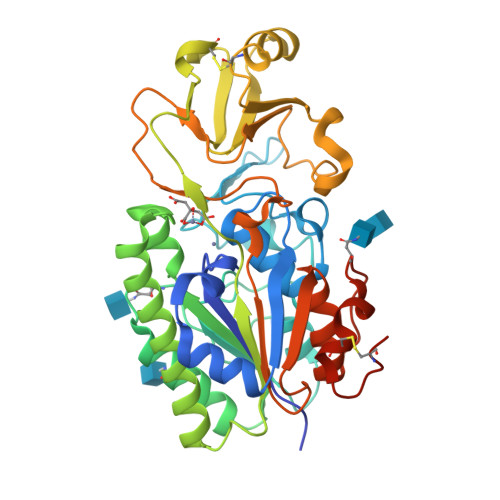
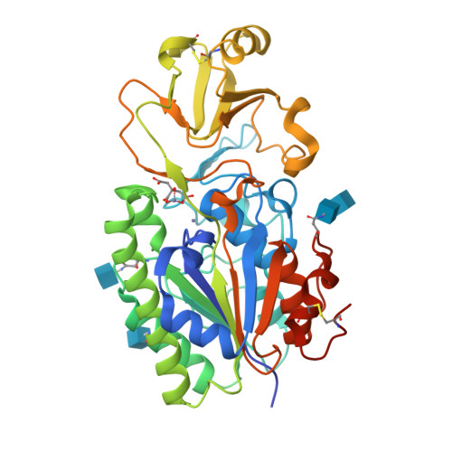
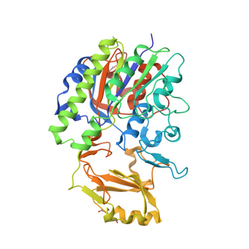
4LR2

Crystal Structure of Human ENPP4 (apo)


Experimental Data Snapshot

  • Method: X-RAY DIFFRACTION
  • Resolution: 1.50 Å
  • R-Value Free: 0.201 
  • R-Value Work: 0.154 
  • R-Value Observed: 0.157 

Starting Model: experimental
View more details

wwPDB Validation   3D Report Full Report


Ligand Structure Quality Assessment 


This is version 2.2 of the entry. See complete history


Literature

Molecular basis of purinergic signal metabolism by ectonucleotide pyrophosphatase/phosphodiesterases 4 and 1 and implications in stroke.

Albright, R.A.Ornstein, D.L.Cao, W.Chang, W.C.Robert, D.Tehan, M.Hoyer, D.Liu, L.Stabach, P.Yang, G.De La Cruz, E.M.Braddock, D.T.

(2014) J Biol Chem 289: 3294-3306

  • DOI: https://doi.org/10.1074/jbc.M113.505867
  • Primary Citation of Related Structures:  
    4LQY, 4LR2

  • PubMed Abstract: 

    NPP4 is a type I extracellular membrane protein on brain vascular endothelium inducing platelet aggregation via the hydrolysis of Ap3A, whereas NPP1 is a type II extracellular membrane protein principally present on the surface of chondrocytes that regulates tissue mineralization. To understand the metabolism of purinergic signals resulting in the physiologic activities of the two enzymes, we report the high resolution crystal structure of human NPP4 and explore the molecular basis of its substrate specificity with NPP1. Both enzymes cleave Ap3A, but only NPP1 can hydrolyze ATP. Comparative structural analysis reveals a tripartite lysine claw in NPP1 that stabilizes the terminal phosphate of ATP, whereas the corresponding region of NPP4 contains features that hinder this binding orientation, thereby inhibiting ATP hydrolysis. Furthermore, we show that NPP1 is unable to induce platelet aggregation at physiologic concentrations reported in human blood, but it could stimulate platelet aggregation if localized at low nanomolar concentrations on vascular endothelium. The combined studies expand our understanding of NPP1 and NPP4 substrate specificity and range and provide a rational mechanism by which polymorphisms in NPP1 confer stroke resistance.


  • Organizational Affiliation

    From the Department of Pathology, Yale University School of Medicine, New Haven, Connecticut 06510.


Macromolecules
Find similar proteins by:  (by identity cutoff)  |  3D Structure
Entity ID: 1
MoleculeChains Sequence LengthOrganismDetailsImage
Bis(5'-adenosyl)-triphosphatase ENPP4401Homo sapiensMutation(s): 0 
Gene Names: ENPP4KIAA0879NPP4
EC: 3.6.1.29
UniProt & NIH Common Fund Data Resources
Find proteins for Q9Y6X5 (Homo sapiens)
Explore Q9Y6X5 
Go to UniProtKB:  Q9Y6X5
PHAROS:  Q9Y6X5
GTEx:  ENSG00000001561 
Entity Groups  
Sequence Clusters30% Identity50% Identity70% Identity90% Identity95% Identity100% Identity
UniProt GroupQ9Y6X5
Glycosylation
Glycosylation Sites: 3Go to GlyGen: Q9Y6X5-1
Sequence Annotations
Expand
  • Reference Sequence
Oligosaccharides

Help

Entity ID: 2
MoleculeChains Length2D Diagram Glycosylation3D Interactions
2-acetamido-2-deoxy-beta-D-glucopyranose-(1-4)-2-acetamido-2-deoxy-beta-D-glucopyranose
B
2N-Glycosylation
Glycosylation Resources
GlyTouCan:  G42666HT
GlyCosmos:  G42666HT
GlyGen:  G42666HT
Experimental Data & Validation

Experimental Data

  • Method: X-RAY DIFFRACTION
  • Resolution: 1.50 Å
  • R-Value Free: 0.201 
  • R-Value Work: 0.154 
  • R-Value Observed: 0.157 
  • Space Group: C 1 2 1
Unit Cell:
Length ( Å )Angle ( ˚ )
a = 181.506α = 90
b = 51.293β = 102.08
c = 53.065γ = 90
Software Package:
Software NamePurpose
DENZOdata reduction
SCALEPACKdata scaling
PHENIXrefinement
PDB_EXTRACTdata extraction

Structure Validation

View Full Validation Report


Ligand Structure Quality Assessment 


Entry History 

Deposition Data

Revision History  (Full details and data files)

  • Version 1.0: 2013-12-18
    Type: Initial release
  • Version 1.1: 2014-02-26
    Changes: Database references
  • Version 1.2: 2017-11-15
    Changes: Refinement description
  • Version 2.0: 2020-07-29
    Type: Remediation
    Reason: Carbohydrate remediation
    Changes: Atomic model, Data collection, Database references, Derived calculations, Structure summary
  • Version 2.1: 2023-09-20
    Changes: Data collection, Database references, Refinement description, Structure summary
  • Version 2.2: 2024-11-20
    Changes: Structure summary