4LO3 | pdb_00004lo3

HA17-HA33-LacNac


Experimental Data Snapshot

  • Method: X-RAY DIFFRACTION
  • Resolution: 2.25 Å
  • R-Value Free: 
    0.213 (Depositor), 0.207 (DCC) 
  • R-Value Work: 
    0.179 (Depositor), 0.175 (DCC) 
  • R-Value Observed: 
    0.181 (Depositor) 

wwPDB Validation 3D Report Full Report

Validation slider image for 4LO3

This is version 2.1 of the entry. See complete history. 

Literature

Structure of a Bimodular Botulinum Neurotoxin Complex Provides Insights into Its Oral Toxicity.

Lee, K., Gu, S., Jin, L., Le, T.T., Cheng, L.W., Strotmeier, J., Kruel, A.M., Yao, G., Perry, K., Rummel, A., Jin, R.

(2013) PLoS Pathog 9: e1003690-e1003690

  • DOI: https://doi.org/10.1371/journal.ppat.1003690
  • Primary Citation Related Structures: 
    4LO0, 4LO1, 4LO2, 4LO3, 4LO4, 4LO5, 4LO6, 4LO7, 4LO8

  • PubMed Abstract: 

    Botulinum neurotoxins (BoNTs) are produced by Clostridium botulinum and cause the fatal disease botulism, a flaccid paralysis of the muscle. BoNTs are released together with several auxiliary proteins as progenitor toxin complexes (PTCs) to become highly potent oral poisons. Here, we report the structure of a ∼760 kDa 14-subunit large PTC of serotype A (L-PTC/A) and reveal insight into its absorption mechanism. Using a combination of X-ray crystallography, electron microscopy, and functional studies, we found that L-PTC/A consists of two structurally and functionally independent sub-complexes. A hetero-dimeric 290 kDa complex protects BoNT, while a hetero-dodecameric 470 kDa complex facilitates its absorption in the harsh environment of the gastrointestinal tract. BoNT absorption is mediated by nine glycan-binding sites on the dodecameric sub-complex that forms multivalent interactions with carbohydrate receptors on intestinal epithelial cells. We identified monosaccharides that blocked oral BoNT intoxication in mice, which suggests a new strategy for the development of preventive countermeasures for BoNTs based on carbohydrate receptor mimicry.


  • Organizational Affiliation: 

    • Department of Physiology and Biophysics, University of California, Irvine, California, United States of America.

Macromolecule Content 

  • Total Structure Weight: 86 kDa 
  • Atom Count: 6,116 
  • Modeled Residue Count: 715 
  • Deposited Residue Count: 739 
  • Unique protein chains: 2

Macromolecules

Find similar proteins by:|  3D Structure
Entity ID: 1
MoleculeChains  Sequence LengthOrganismDetailsImage
HA-33A,
C [auth B]
296Clostridium botulinumMutation(s): 0 
Gene Names: HA-33, ha33, ha34
UniProt
Find proteins for Q45871 (Clostridium botulinum)
Explore Q45871 
Go to UniProtKB:  Q45871
Entity Groups
Sequence Clusters30% Identity50% Identity70% Identity90% Identity95% Identity100% Identity
UniProt GroupQ45871
Sequence Annotations
Expand
Reference Sequence
Find similar proteins by:|  3D Structure
Entity ID: 2
MoleculeChains  Sequence LengthOrganismDetailsImage
HA-17B [auth C]147Clostridium botulinumMutation(s): 0 
Gene Names: ha17, HA-17
UniProt
Find proteins for Q45878 (Clostridium botulinum)
Explore Q45878 
Go to UniProtKB:  Q45878
Entity Groups
Sequence Clusters30% Identity50% Identity70% Identity90% Identity95% Identity100% Identity
UniProt GroupQ45878
Sequence Annotations
Expand
Reference Sequence

Oligosaccharides

Help  
Entity ID: 3
MoleculeChains Length2D Diagram GlycosylationD Interactions
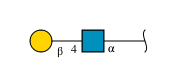
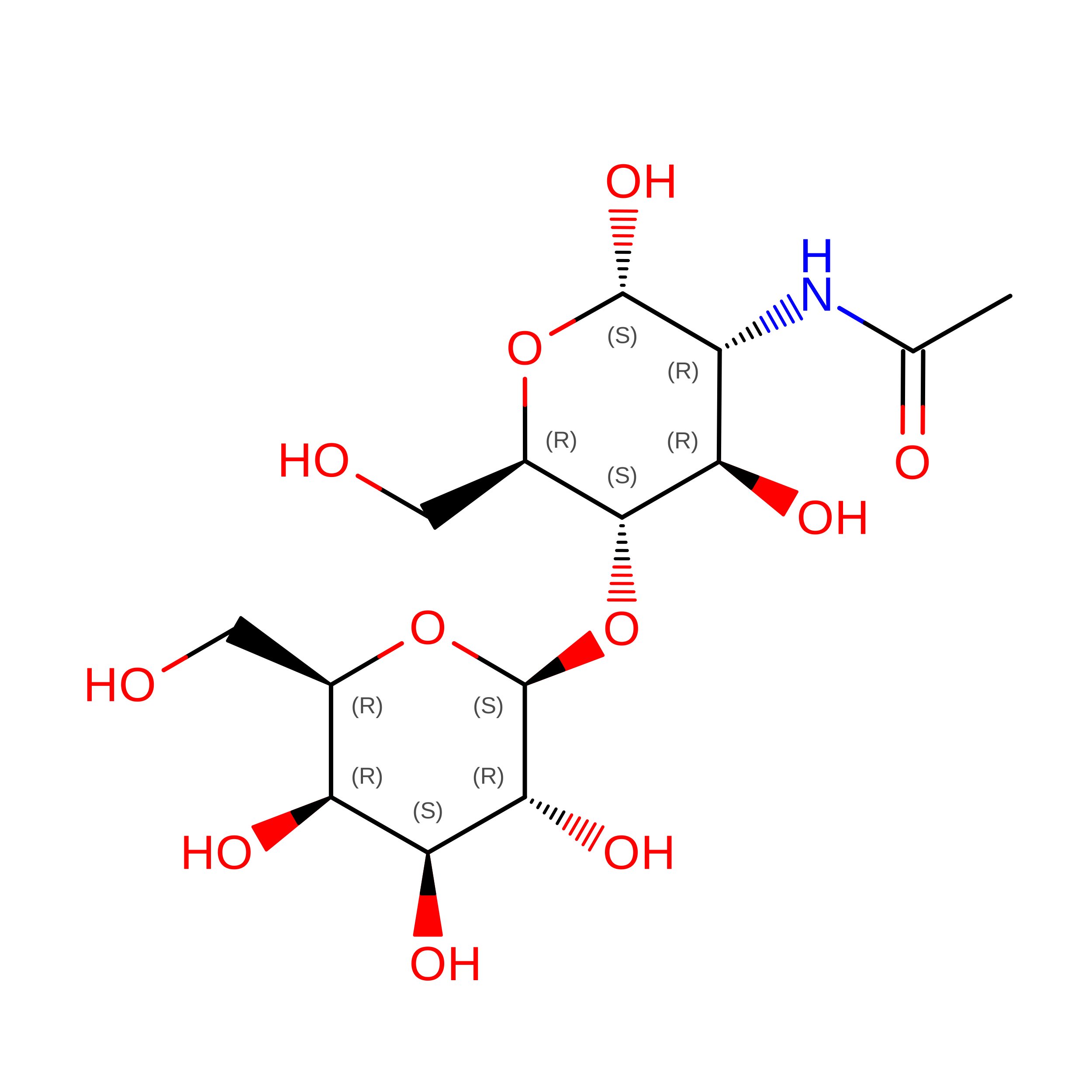
beta-D-galactopyranose-(1-4)-2-acetamido-2-deoxy-alpha-D-glucopyranose
D, E
2N/A
Glycosylation Resources
GlyTouCan: G79333MY
GlyCosmos: G79333MY
GlyGen: G79333MY

Biologically Interesting Molecules (External Reference) 

1 Unique

Experimental Data & Validation

Experimental Data

  • Method: X-RAY DIFFRACTION
  • Resolution: 2.25 Å
  • R-Value Free:  0.213 (Depositor), 0.207 (DCC) 
  • R-Value Work:  0.179 (Depositor), 0.175 (DCC) 
  • R-Value Observed: 0.181 (Depositor) 
Space Group: C 2 2 21
Unit Cell:
Length ( Å )Angle ( ˚ )
a = 107.216α = 90
b = 118.803β = 90
c = 162.536γ = 90
Software Package:
Software NamePurpose
Blu-Icedata collection
PHASESphasing
PHENIXrefinement
HKL-2000data reduction
HKL-2000data scaling

Structure Validation

View Full Validation Report


Entry History 

Deposition Data

Revision History  (Full details and data files)

  • Version 1.0: 2013-10-30
    Type: Initial release
  • Version 2.0: 2020-07-29
    Type: Remediation
    Reason: Carbohydrate remediation
    Changes: Advisory, Atomic model, Data collection, Database references, Derived calculations, Structure summary
  • Version 2.1: 2024-02-28
    Changes: Advisory, Data collection, Database references, Structure summary