4LMX | pdb_00004lmx

Light harvesting complex PE555 from the cryptophyte Hemiselmis andersenii CCMP644


Experimental Data Snapshot

  • Method: X-RAY DIFFRACTION
  • Resolution: 1.80 Å
  • R-Value Free: 
    0.214 (Depositor), 0.211 (DCC) 
  • R-Value Work: 
    0.158 (Depositor), 0.155 (DCC) 
  • R-Value Observed: 
    0.161 (Depositor) 

Starting Model: experimental
View more details

wwPDB Validation 3D Report Full Report

Validation slider image for 4LMX

Ligand Structure Quality Assessment 


This is version 1.3 of the entry. See complete history

Literature

Single-residue insertion switches the quaternary structure and exciton states of cryptophyte light-harvesting proteins.

Harrop, S.J.Wilk, K.E.Dinshaw, R.Collini, E.Mirkovic, T.Teng, C.Y.Oblinsky, D.G.Green, B.R.Hoef-Emden, K.Hiller, R.G.Scholes, G.D.Curmi, P.M.

(2014) Proc Natl Acad Sci U S A 111: E2666-E2675

  • DOI: https://doi.org/10.1073/pnas.1402538111
  • Primary Citation Related Structures: 
    4LM6, 4LMS, 4LMX

  • PubMed Abstract: 

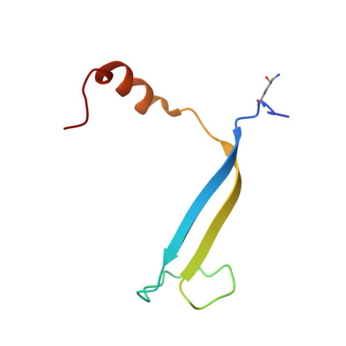
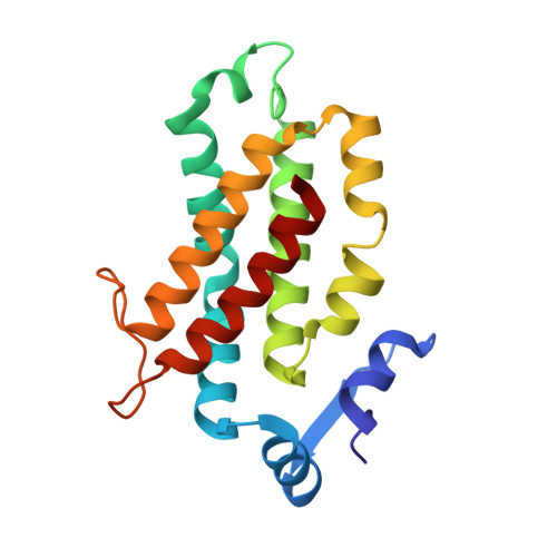
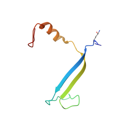
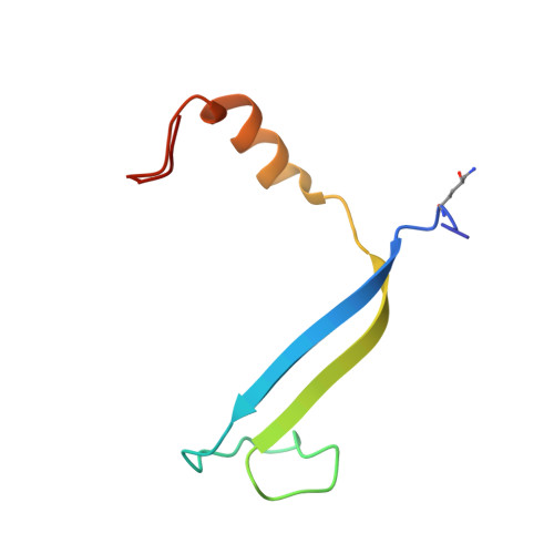
    Observation of coherent oscillations in the 2D electronic spectra (2D ES) of photosynthetic proteins has led researchers to ask whether nontrivial quantum phenomena are biologically significant. Coherent oscillations have been reported for the soluble light-harvesting phycobiliprotein (PBP) antenna isolated from cryptophyte algae. To probe the link between spectral properties and protein structure, we determined crystal structures of three PBP light-harvesting complexes isolated from different species. Each PBP is a dimer of αβ subunits in which the structure of the αβ monomer is conserved. However, we discovered two dramatically distinct quaternary conformations, one of which is specific to the genus Hemiselmis. Because of steric effects emerging from the insertion of a single amino acid, the two αβ monomers are rotated by ∼73° to an "open" configuration in contrast to the "closed" configuration of other cryptophyte PBPs. This structural change is significant for the light-harvesting function because it disrupts the strong excitonic coupling between two central chromophores in the closed form. The 2D ES show marked cross-peak oscillations assigned to electronic and vibrational coherences in the closed-form PC645. However, such features appear to be reduced, or perhaps absent, in the open structures. Thus cryptophytes have evolved a structural switch controlled by an amino acid insertion to modulate excitonic interactions and therefore the mechanisms used for light harvesting.


  • Organizational Affiliation
    • School of Physics, The University of New South Wales, Sydney, NSW 2052, Australia;

Macromolecule Content 

  • Total Structure Weight: 166.48 kDa 
  • Atom Count: 14,008 
  • Modeled Residue Count: 1,473 
  • Deposited Residue Count: 1,487 
  • Unique protein chains: 4

Macromolecules

Find similar proteins by:|  3D Structure
Entity ID: 1
MoleculeChains  Sequence LengthOrganismDetailsImage
cryptophyte phycoerythrin (alpha-2 chain)62Hemiselmis anderseniiMutation(s): 0 
UniProt
Find proteins for U5TBJ3 (Hemiselmis andersenii)
Explore U5TBJ3 
Go to UniProtKB:  U5TBJ3
Entity Groups
Sequence Clusters30% Identity50% Identity70% Identity90% Identity95% Identity100% Identity
UniProt GroupU5TBJ3
Sequence Annotations
Expand
Reference Sequence
Find similar proteins by:|  3D Structure
Entity ID: 2
MoleculeChains  Sequence LengthOrganismDetailsImage
cryptophyte phycoerythrin (beta chain)
B, D, F, H, J
B, D, F, H, J, L
177Hemiselmis anderseniiMutation(s): 0 
UniProt
Find proteins for U5T8W0 (Hemiselmis andersenii)
Explore U5T8W0 
Go to UniProtKB:  U5T8W0
Entity Groups
Sequence Clusters30% Identity50% Identity70% Identity90% Identity95% Identity100% Identity
UniProt GroupU5T8W0
Sequence Annotations
Expand
Reference Sequence
Find similar proteins by:|  3D Structure
Entity ID: 3
MoleculeChains  Sequence LengthOrganismDetailsImage
cryptophyte phycoerythrin (alpha-1 chain)67Hemiselmis anderseniiMutation(s): 0 
UniProt
Find proteins for U5TBU5 (Hemiselmis andersenii)
Explore U5TBU5 
Go to UniProtKB:  U5TBU5
Entity Groups
Sequence Clusters30% Identity50% Identity70% Identity90% Identity95% Identity100% Identity
UniProt GroupU5TBU5
Sequence Annotations
Expand
Reference Sequence
Find similar proteins by:|  3D Structure
Entity ID: 4
MoleculeChains  Sequence LengthOrganismDetailsImage
cryptophyte phycoerythrin (alpha-1/alpha-2 chain)
E, G, I, K
67Hemiselmis anderseniiMutation(s): 7 
UniProt
Find proteins for U5TBJ3 (Hemiselmis andersenii)
Explore U5TBJ3 
Go to UniProtKB:  U5TBJ3
Entity Groups
Sequence Clusters30% Identity50% Identity70% Identity90% Identity95% Identity100% Identity
UniProt GroupU5TBJ3
Sequence Annotations
Expand
Reference Sequence

Small Molecules

Ligands 2 Unique
IDChains Name / Formula / InChI Key2D Diagram3D Interactions
PEB

Query on PEB


Download:Ideal Coordinates CCD File
AA [auth H]
BA [auth H]
CA [auth I]
EA [auth J]
FA [auth J]
AA [auth H],
BA [auth H],
CA [auth I],
EA [auth J],
FA [auth J],
GA [auth K],
IA [auth L],
JA [auth L],
M [auth A],
O [auth B],
P [auth B],
Q [auth C],
S [auth D],
T [auth D],
U [auth E],
W [auth F],
X [auth F],
Y [auth G]
PHYCOERYTHROBILIN
C33 H40 N4 O6
NKCBCVIFPXGHAV-WAVSMFBNSA-N
DBV

Query on DBV


Download:Ideal Coordinates CCD File
DA [auth J]
HA [auth L]
N [auth B]
R [auth D]
V [auth F]
DA [auth J],
HA [auth L],
N [auth B],
R [auth D],
V [auth F],
Z [auth H]
15,16-DIHYDROBILIVERDIN
C33 H36 N4 O6
ZQHDSLZHMAUUQK-ZTYGKHTCSA-N
Modified Residues  1 Unique
IDChains TypeFormula2D DiagramParent
LYZ
Query on LYZ
A
L-PEPTIDE LINKINGC6 H14 N2 O3LYS

Experimental Data & Validation

Experimental Data

  • Method: X-RAY DIFFRACTION
  • Resolution: 1.80 Å
  • R-Value Free:  0.214 (Depositor), 0.211 (DCC) 
  • R-Value Work:  0.158 (Depositor), 0.155 (DCC) 
  • R-Value Observed: 0.161 (Depositor) 
Space Group: P 1 21 1
Unit Cell:
Length ( Å )Angle ( ˚ )
a = 66.073α = 90
b = 76.741β = 92.86
c = 142.593γ = 90
Software Package:
Software NamePurpose
Blu-Icedata collection
PHASERphasing
PHENIXrefinement
XDSdata reduction
SCALAdata scaling

Structure Validation

View Full Validation Report


Ligand Structure Quality Assessment 


Entry History 

Deposition Data

Revision History  (Full details and data files)

  • Version 1.0: 2014-06-18
    Type: Initial release
  • Version 1.1: 2014-07-16
    Changes: Database references
  • Version 1.2: 2023-09-20
    Changes: Data collection, Database references, Derived calculations, Refinement description
  • Version 1.3: 2024-11-27
    Changes: Structure summary