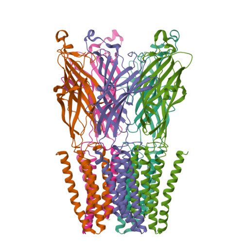
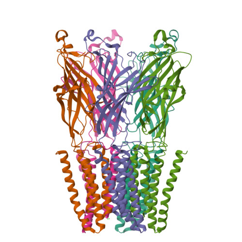
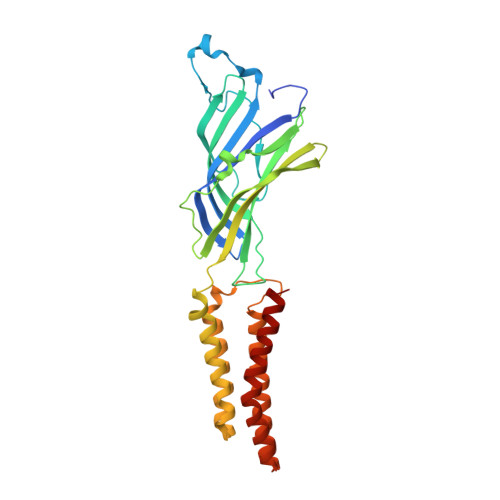
4LMJ

GLIC Liganded-closed-channel Conformation, Mutant T25'A


Experimental Data Snapshot

  • Method: X-RAY DIFFRACTION
  • Resolution: 3.44 Å
  • R-Value Free: 
    0.255 (Depositor), 0.250 (DCC) 
  • R-Value Work: 
    0.227 (Depositor), 0.230 (DCC) 
  • R-Value Observed: 
    0.228 (Depositor) 

Starting Model: experimental
View more details

wwPDB Validation   3D Report Full Report


Ligand Structure Quality Assessment 

Created with Raphaël 2.3.0Worse 01 BetterLigand structure goodness of fit to experimental dataBest fitted LMTClick on this verticalbar to view details

This is version 1.3 of the entry. See complete history


Literature

Gating of the proton-gated ion channel from Gloeobacter violaceus at pH 4 as revealed by X-ray crystallography.

Gonzalez-Gutierrez, G.Cuello, L.G.Nair, S.K.Grosman, C.

(2013) Proc Natl Acad Sci U S A 110: 18716-18721

  • DOI: https://doi.org/10.1073/pnas.1313156110
  • Primary Citation of Related Structures:  
    4LMJ, 4LMK, 4LML

  • PubMed Abstract: 

    Cryoelectron microscopy and X-ray crystallography have recently been used to generate structural models that likely represent the unliganded closed-channel conformation and the fully liganded open-channel conformation of different members of the nicotinic-receptor superfamily. To characterize the structure of the closed-channel conformation in its liganded state, we identified a number of positions in the loop between transmembrane segments 2 (M2) and 3 (M3) of a proton-gated ortholog from the bacterium Gloeobacter violaceus (GLIC) where mutations to alanine reduce the liganded-gating equilibrium constant, and solved the crystal structures of two such mutants (T25'A and Y27'A) at pH ~4.0. At the level of backbone atoms, the liganded closed-channel model presented here differs from the liganded open-channel structure of GLIC in the pre-M1 linker, the M3-M4 loop, and much more prominently, in the extracellular half of the pore lining, where the more pronounced tilt of the closed-channel M2 α-helices toward the pore's long axis narrows the permeation pathway. On the other hand, no differences between the liganded closed-channel and open-channel models could be detected at the level of the extracellular domain, where conformational changes are expected to underlie the low-to-high proton-affinity switch that drives gating of proton-bound channels. Thus, the liganded closed-channel model is nearly indistinguishable from the recently described "locally closed" structure. However, because cross-linking strategies (which could have stabilized unstable conformations) and mutations involving ionizable side chains (which could have affected proton-gated channel activation) were purposely avoided, we favor the notion that this structure represents one of the end states of liganded gating rather than an unstable intermediate.


  • Organizational Affiliation

    Department of Molecular and Integrative Physiology, Center for Biophysics and Computational Biology, Department of Biochemistry, Institute for Genomic Biology, and Neuroscience Program, University of Illinois at Urbana-Champaign, Urbana, IL 61801.


Macromolecules
Find similar proteins by:  (by identity cutoff)  |  3D Structure
Entity ID: 1
MoleculeChains Sequence LengthOrganismDetailsImage
Proton-gated ion channelA [auth E],
B,
C,
D [auth A],
E [auth D]
318Gloeobacter violaceusMutation(s): 1 
Gene Names: glvIglr4197
Membrane Entity: Yes 
UniProt
Find proteins for Q7NDN8 (Gloeobacter violaceus (strain ATCC 29082 / PCC 7421))
Explore Q7NDN8 
Go to UniProtKB:  Q7NDN8
Entity Groups  
Sequence Clusters30% Identity50% Identity70% Identity90% Identity95% Identity100% Identity
UniProt GroupQ7NDN8
Sequence Annotations
Expand
  • Reference Sequence
Small Molecules
Ligands 3 Unique
IDChains Name / Formula / InChI Key2D Diagram3D Interactions
LMT
Query on LMT

Download Ideal Coordinates CCD File 
G [auth E]DODECYL-BETA-D-MALTOSIDE
C24 H46 O11
NLEBIOOXCVAHBD-QKMCSOCLSA-N
CL
Query on CL

Download Ideal Coordinates CCD File 
F [auth E],
I [auth B],
L [auth C],
N [auth A],
P [auth D]
CHLORIDE ION
Cl
VEXZGXHMUGYJMC-UHFFFAOYSA-M
NA
Query on NA

Download Ideal Coordinates CCD File 
H [auth B],
J [auth B],
K [auth C],
M [auth A],
O [auth D]
SODIUM ION
Na
FKNQFGJONOIPTF-UHFFFAOYSA-N
Experimental Data & Validation

Experimental Data

  • Method: X-RAY DIFFRACTION
  • Resolution: 3.44 Å
  • R-Value Free:  0.255 (Depositor), 0.250 (DCC) 
  • R-Value Work:  0.227 (Depositor), 0.230 (DCC) 
  • R-Value Observed: 0.228 (Depositor) 
Space Group: C 1 2 1
Unit Cell:
Length ( Å )Angle ( ˚ )
a = 181.12α = 90
b = 134.32β = 101.96
c = 159.89γ = 90
Software Package:
Software NamePurpose
PHENIXrefinement
PDB_EXTRACTdata extraction
HKL-2000data reduction
HKL-2000data scaling
PHENIXphasing

Structure Validation

View Full Validation Report


Ligand Structure Quality Assessment 

Created with Raphaël 2.3.0Worse 01 BetterLigand structure goodness of fit to experimental dataBest fitted LMTClick on this verticalbar to view details

Entry History 

Deposition Data

Revision History  (Full details and data files)

  • Version 1.0: 2013-10-30
    Type: Initial release
  • Version 1.1: 2013-11-13
    Changes: Database references
  • Version 1.2: 2013-11-27
    Changes: Database references
  • Version 1.3: 2023-09-20
    Changes: Data collection, Database references, Derived calculations, Refinement description