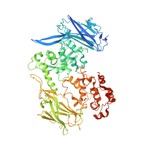
Insights into electron transfer at the microbe-mineral interface: the X-ray crystal structures of Shewanella oneidensis MtrC and OmcA
Edwards, M.J., Baiden, N., Clarke, T.A., Richardson, D.J.(2014) Febs Letters
Experimental Data Snapshot
(2014) Febs Letters
Entity ID: 1 | |||||
|---|---|---|---|---|---|
| Molecule | Chains | Sequence Length | Organism | Details | Image |
| Extracellular iron oxide respiratory system surface decaheme cytochrome c component OmcA | 760 | Shewanella oneidensis MR-1 | Mutation(s): 0 Gene Names: omcA, SO_1779 |  | |
UniProt | |||||
Find proteins for Q8EG33 (Shewanella oneidensis (strain ATCC 700550 / JCM 31522 / CIP 106686 / LMG 19005 / NCIMB 14063 / MR-1)) Explore Q8EG33 Go to UniProtKB: Q8EG33 | |||||
Entity Groups | |||||
| Sequence Clusters | 30% Identity50% Identity70% Identity90% Identity95% Identity100% Identity | ||||
| UniProt Group | Q8EG33 | ||||
Sequence AnnotationsExpand | |||||
| |||||
| Ligands 3 Unique | |||||
|---|---|---|---|---|---|
| ID | Chains | Name / Formula / InChI Key | 2D Diagram | 3D Interactions | |
| HEC Query on HEC | AA [auth B] AB [auth D] BA [auth B] BB [auth D] CA [auth B] | HEME C C34 H34 Fe N4 O4 HXQIYSLZKNYNMH-LJNAALQVSA-N |  | ||
| DMS Query on DMS | HA [auth B] IA [auth B] JA [auth B] MB [auth D] NB [auth D] | DIMETHYL SULFOXIDE C2 H6 O S IAZDPXIOMUYVGZ-UHFFFAOYSA-N |  | ||
| CA Query on CA | FA [auth B] GA [auth B] KB [auth D] LB [auth D] O [auth A] | CALCIUM ION Ca BHPQYMZQTOCNFJ-UHFFFAOYSA-N |  | ||
| Length ( Å ) | Angle ( ˚ ) |
|---|---|
| a = 92.64 | α = 90 |
| b = 245.38 | β = 97.89 |
| c = 135.63 | γ = 90 |
| Software Name | Purpose |
|---|---|
| XDS | data scaling |
| SHELXS | phasing |
| PHENIX | refinement |
| xia2 | data reduction |
| XDS | data reduction |
| SCALA | data scaling |